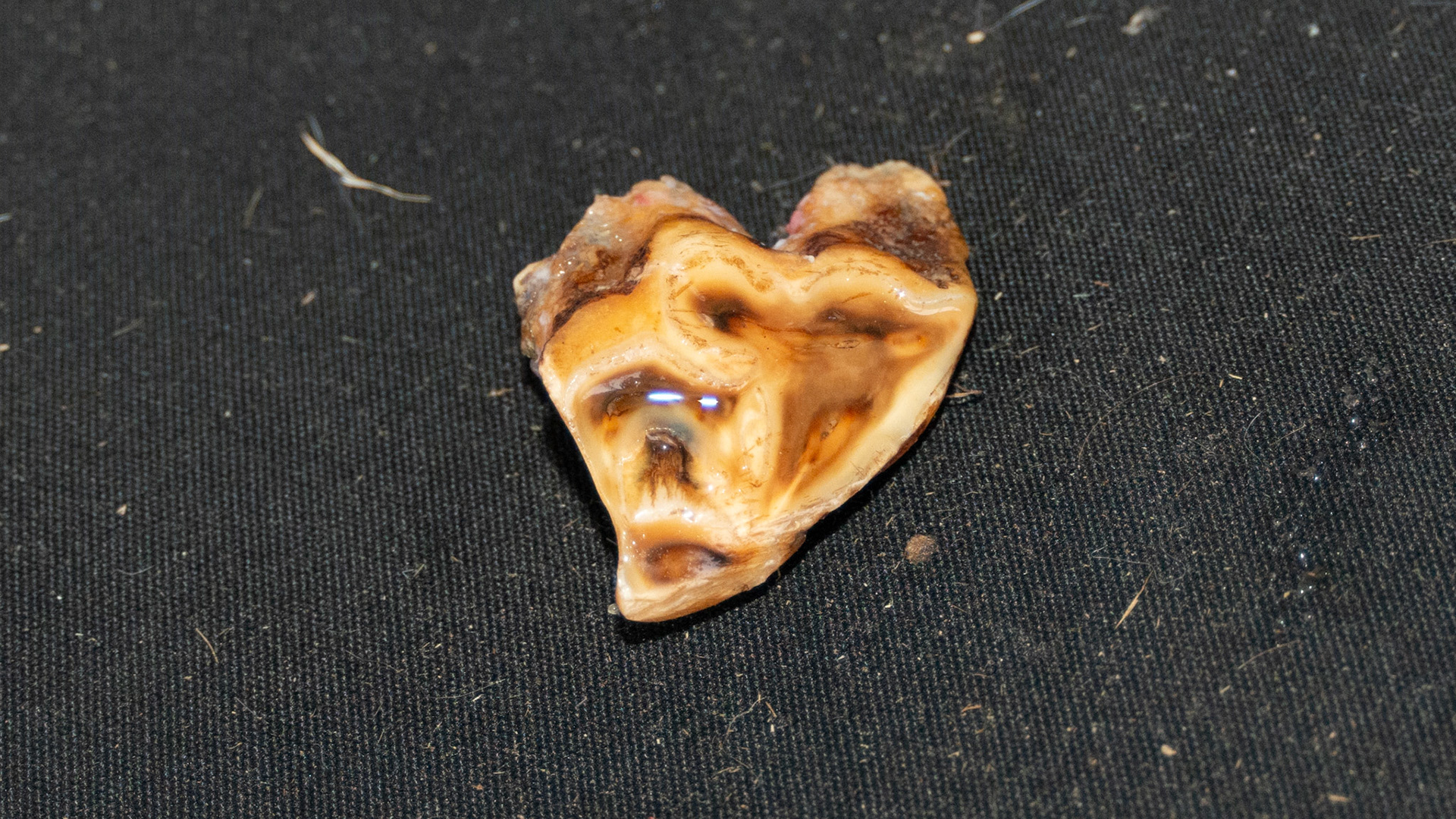
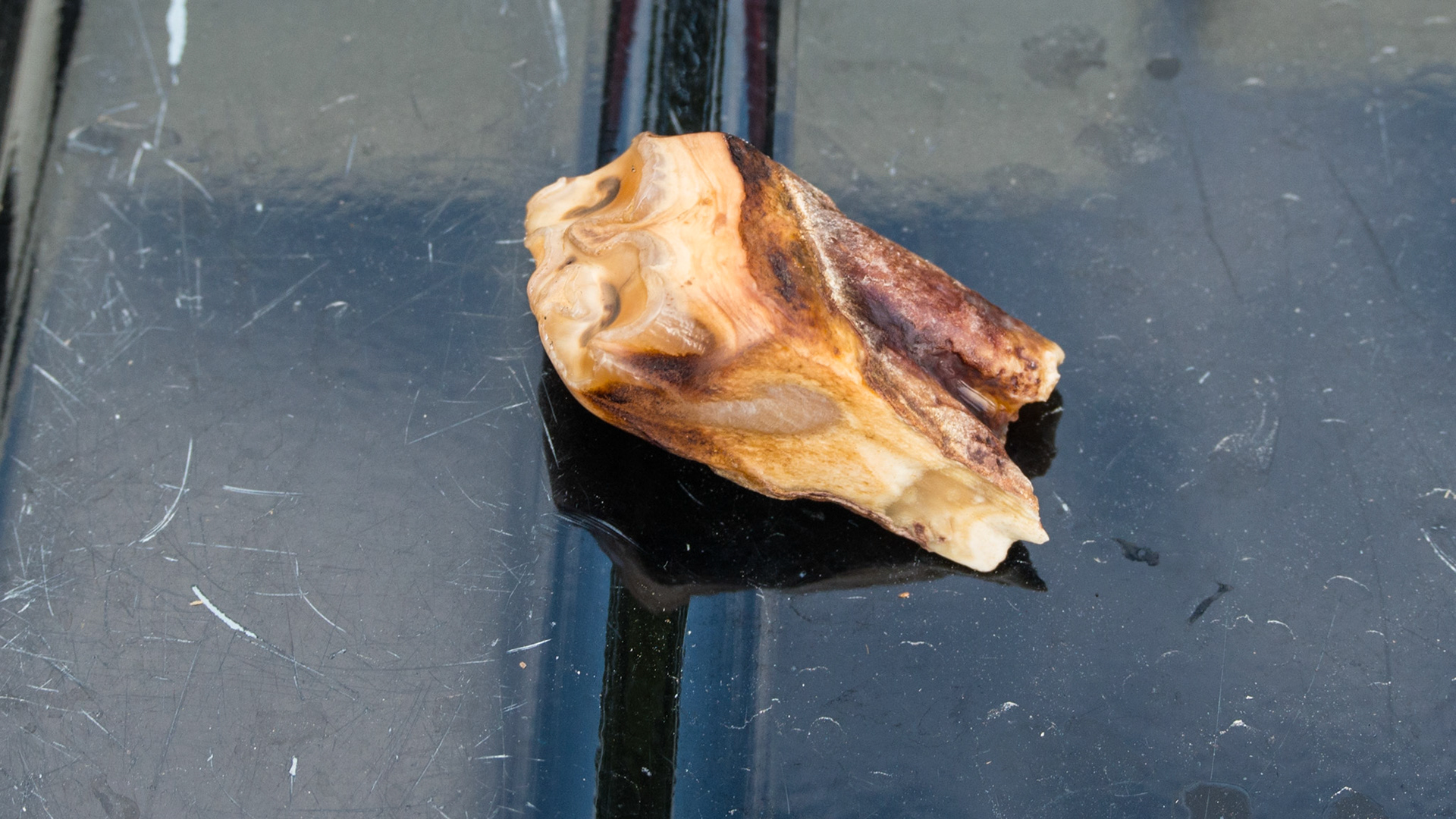
End stage, necrotic #206 in a 23 year old horse.
End stage, necrotic #206 in a 23 year old horse.
End stage, necrotic #206 in a 23 year old horse.
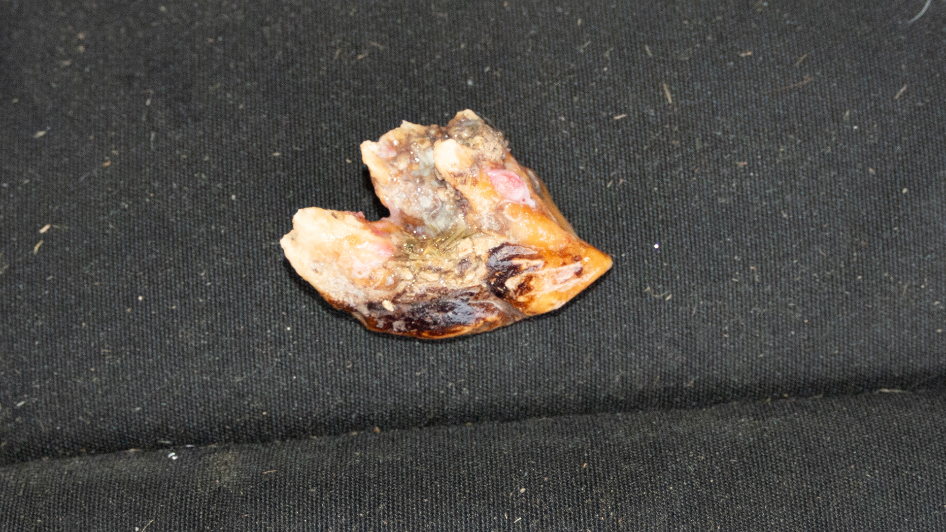
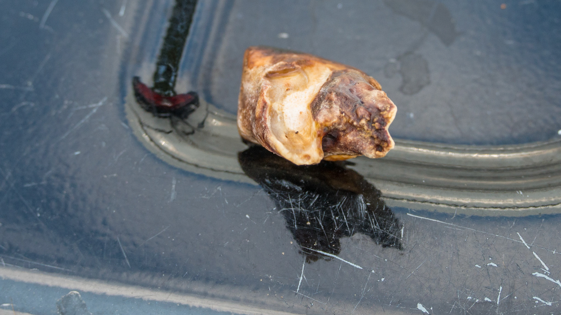
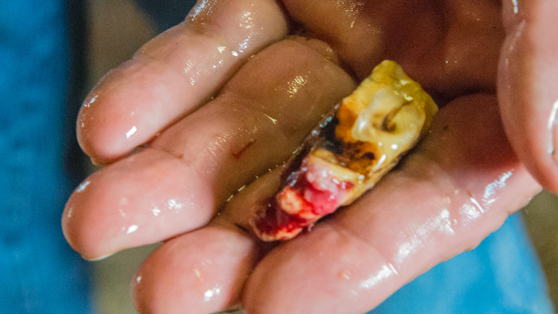
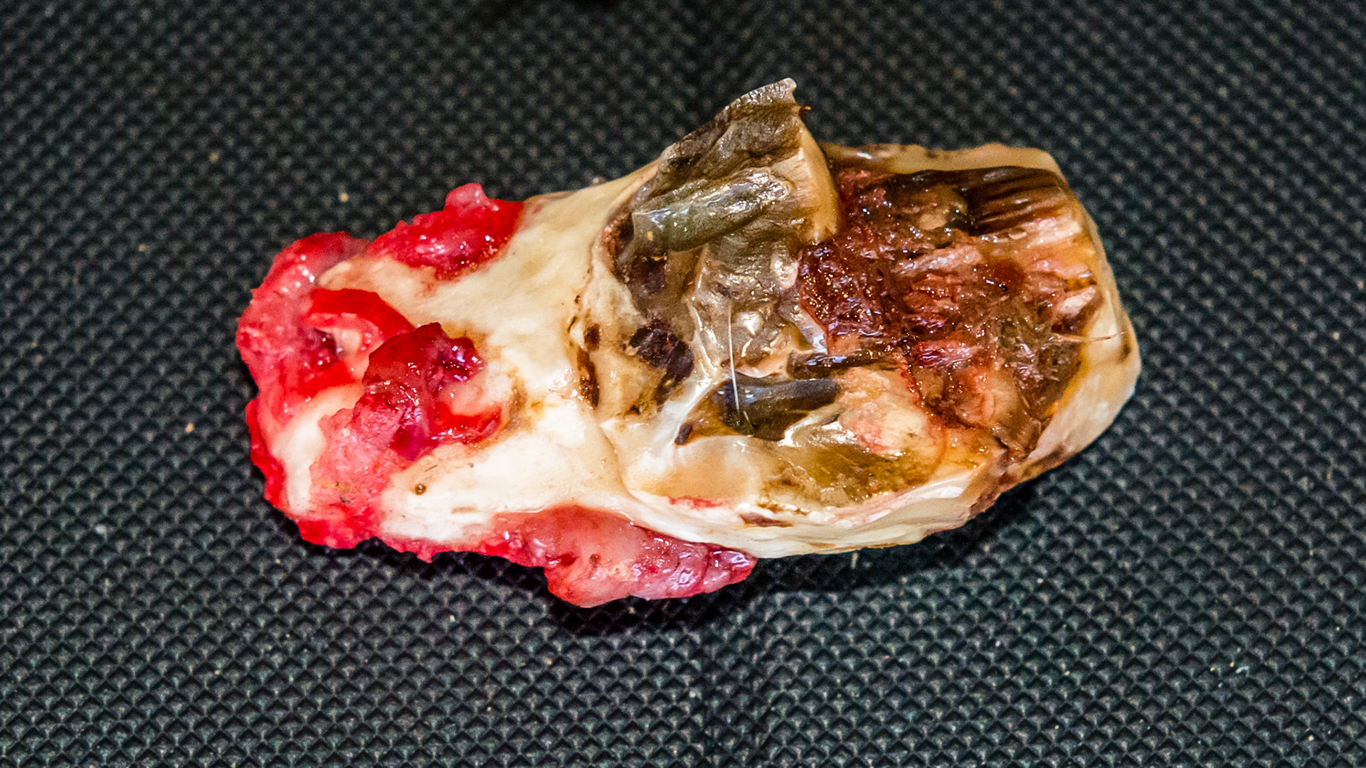
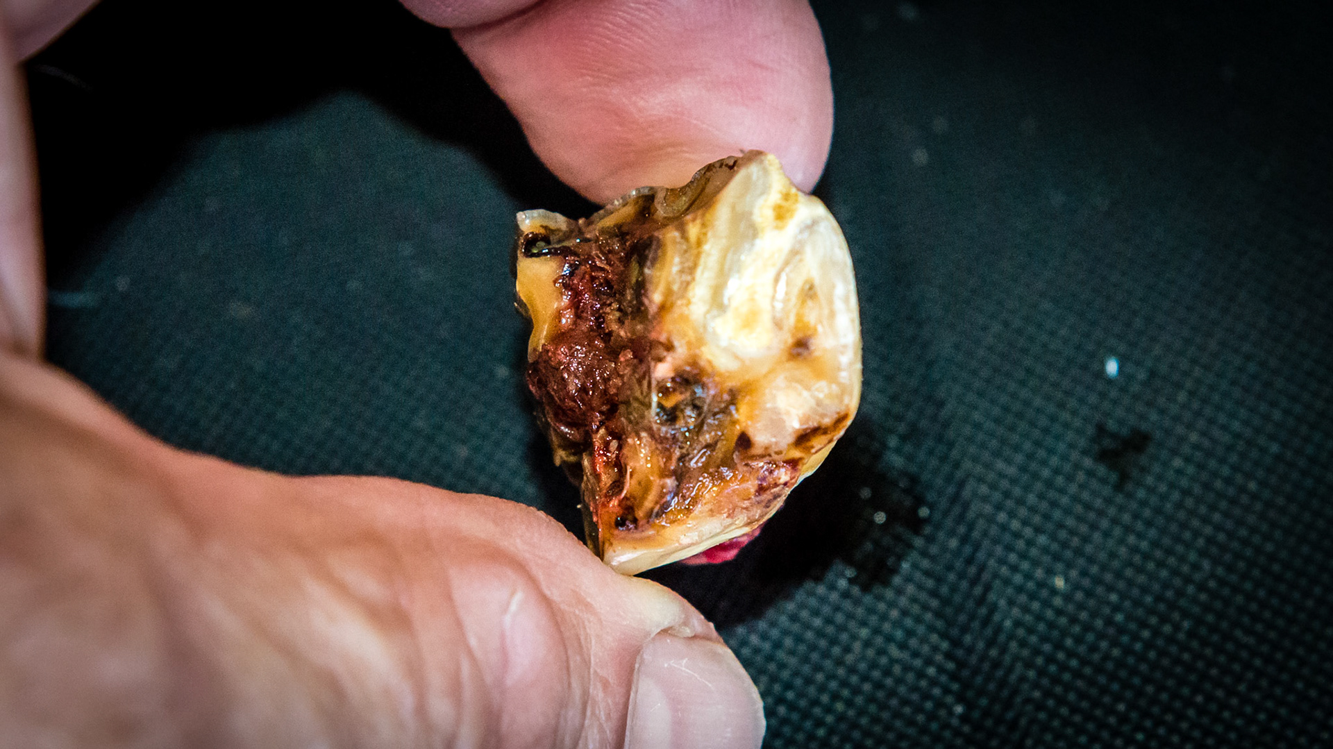
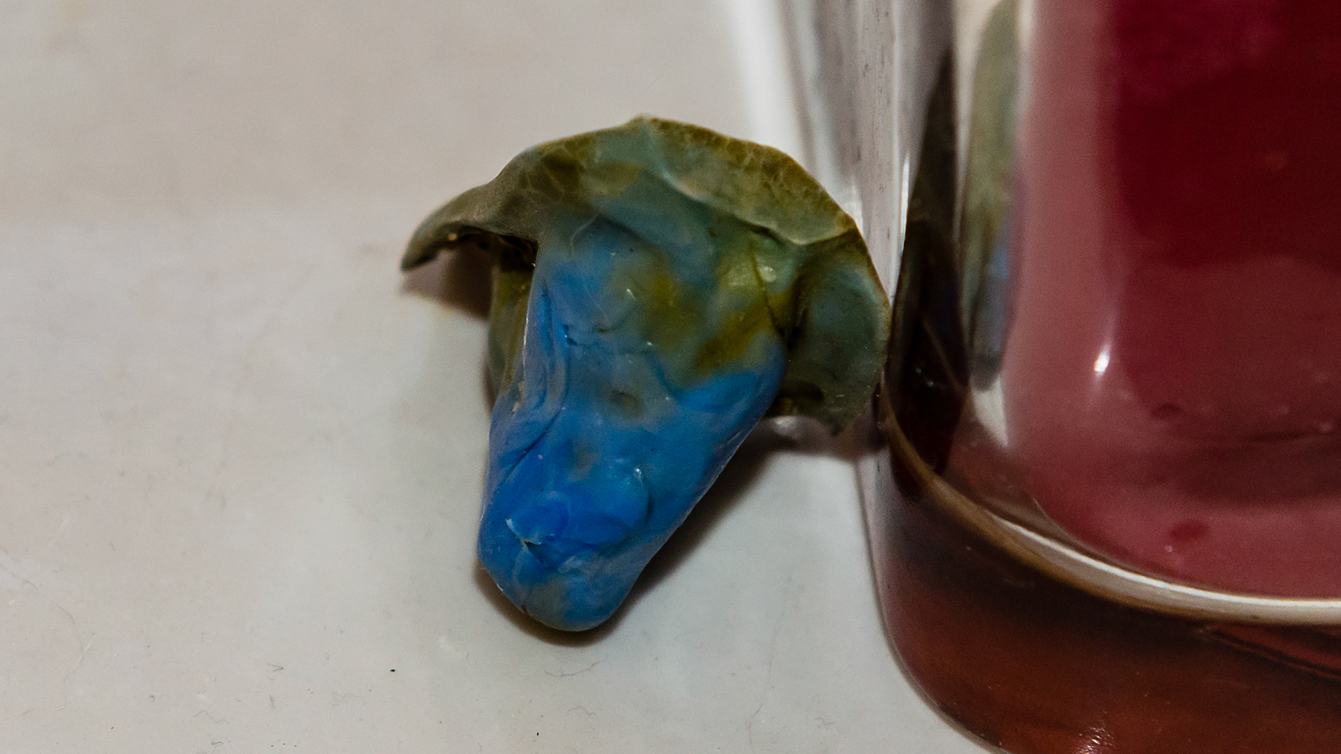
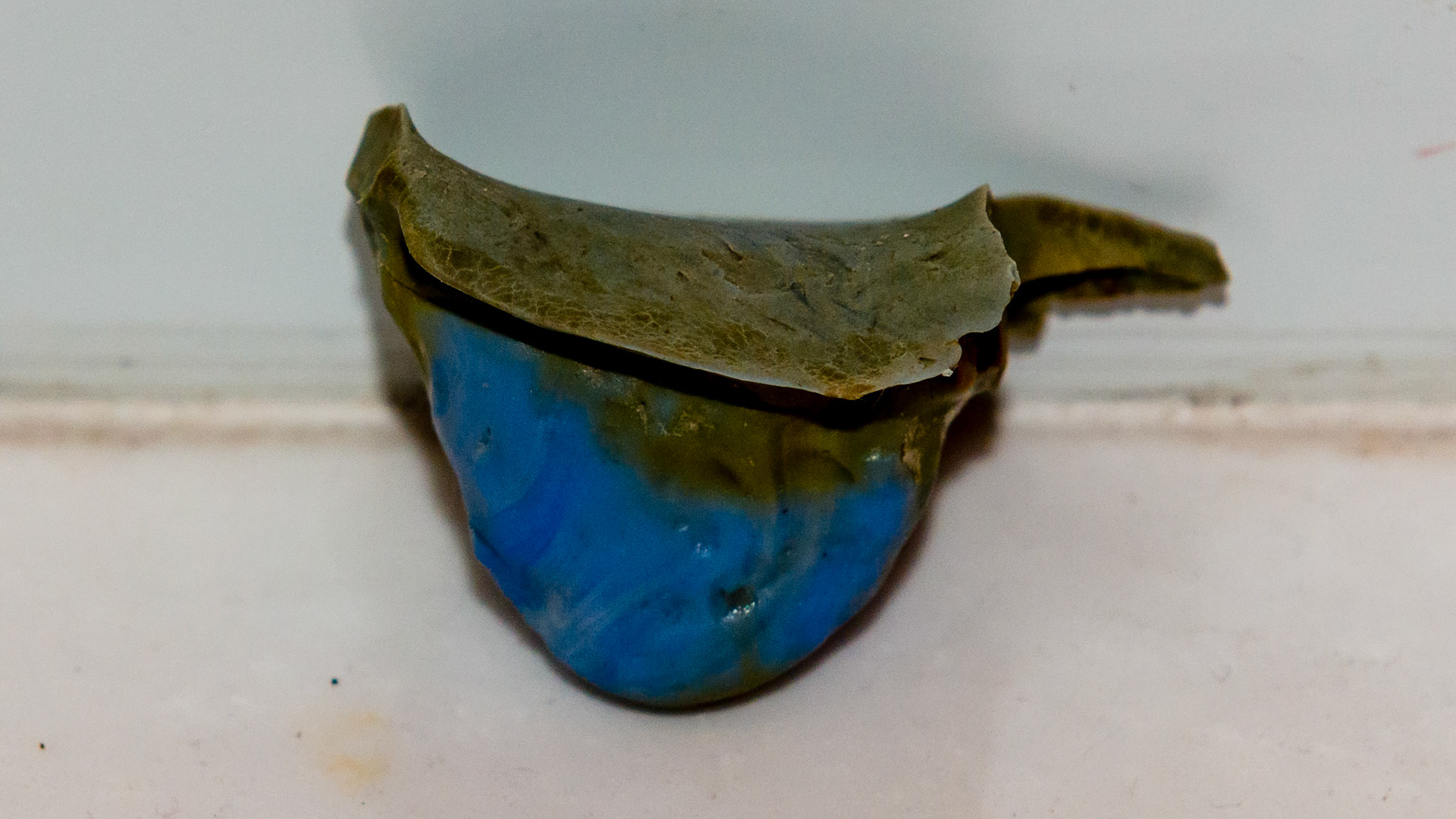
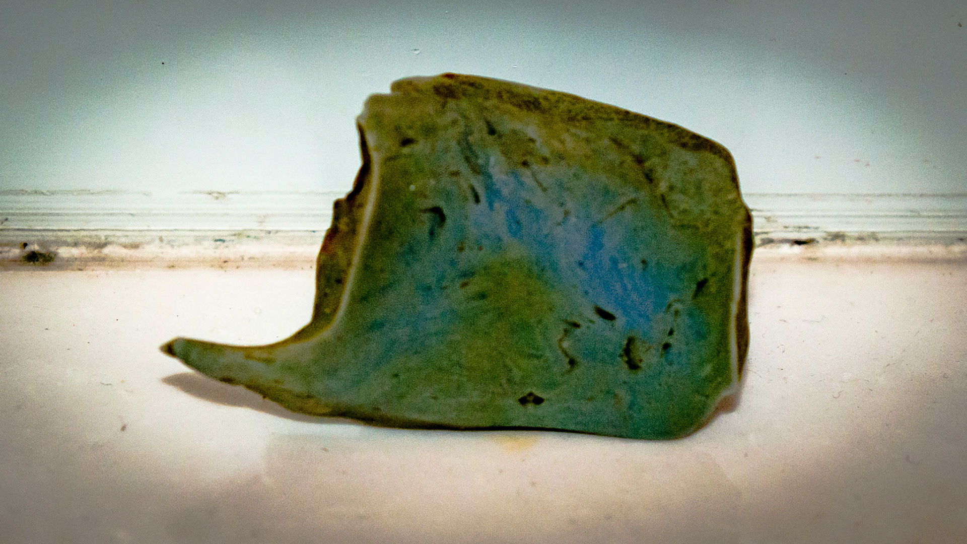
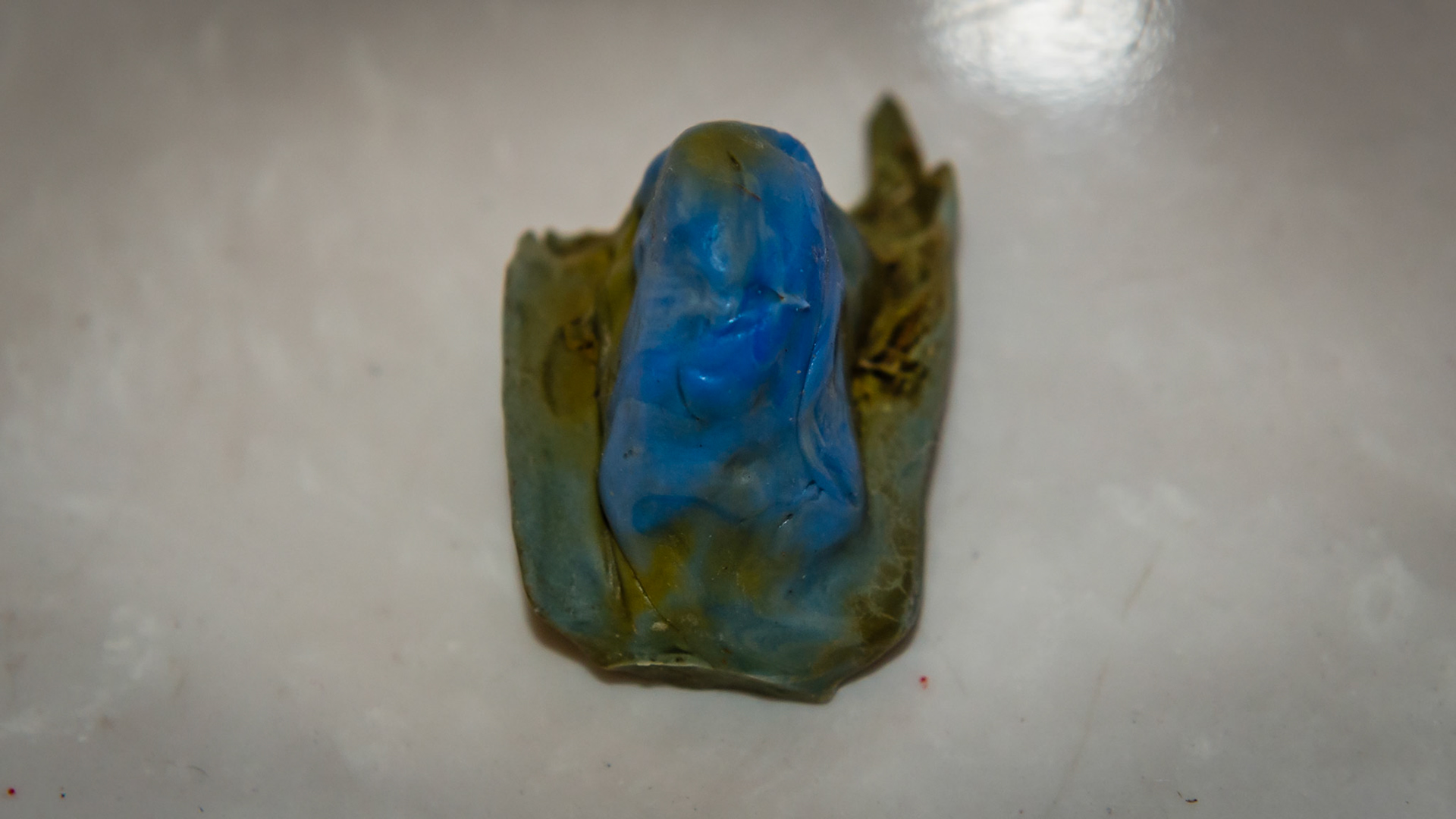
Extracted end stage cheek tooth
Extracted end stage cheek tooth
Extracted end stage cheek tooth
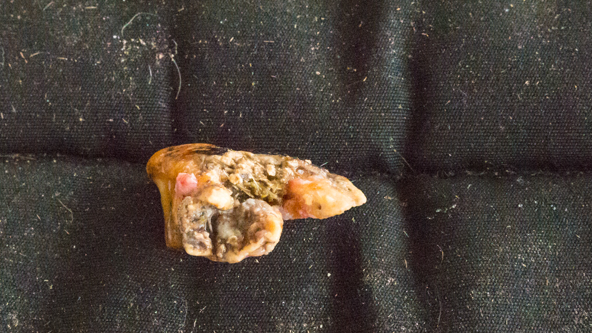
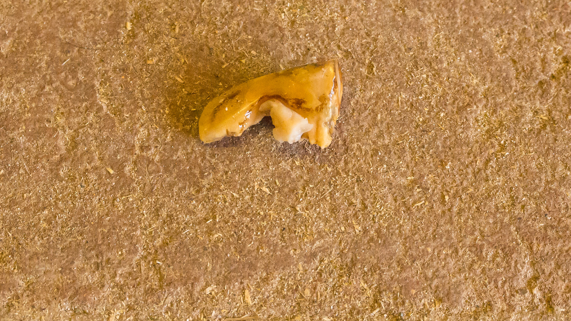
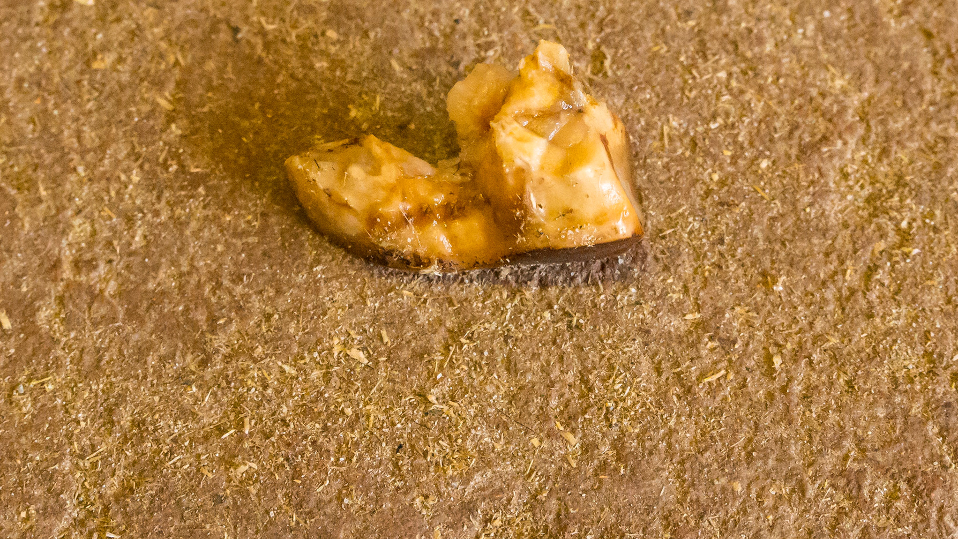
End stage #406 from a 28 year old
End stage #406 from a 28 year old
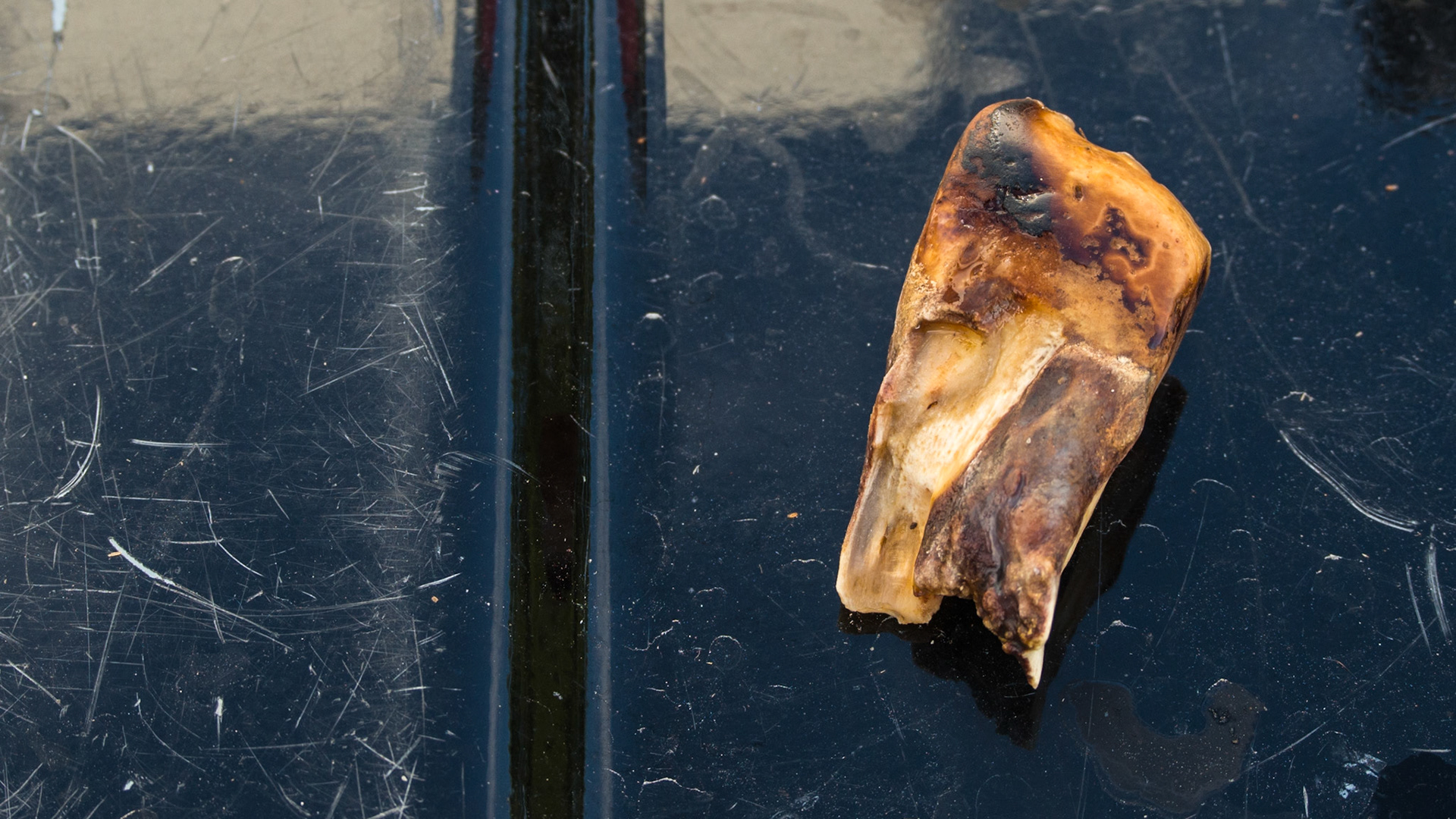
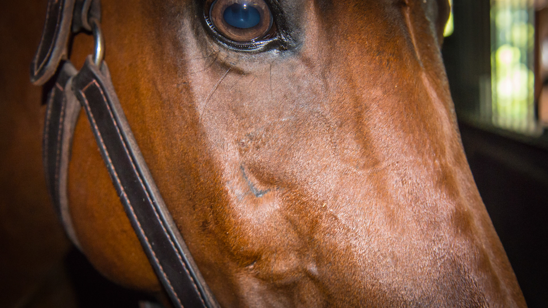
Tooth extraction #109 performed about 2 years ago.
Tooth extraction #109 performed about 2 years ago.
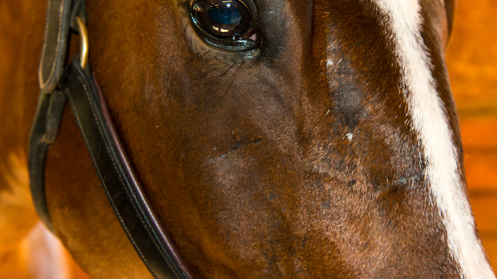
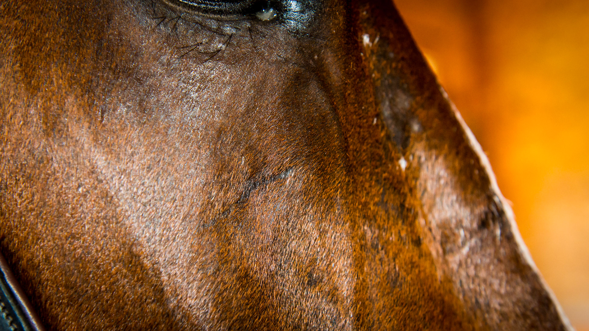
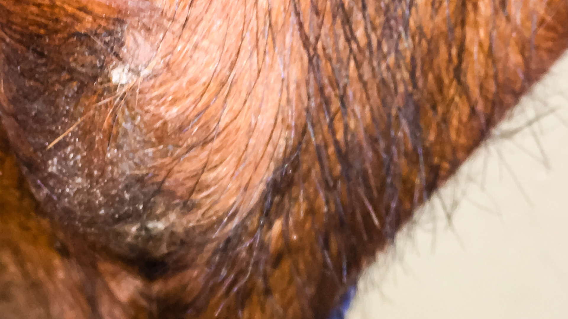
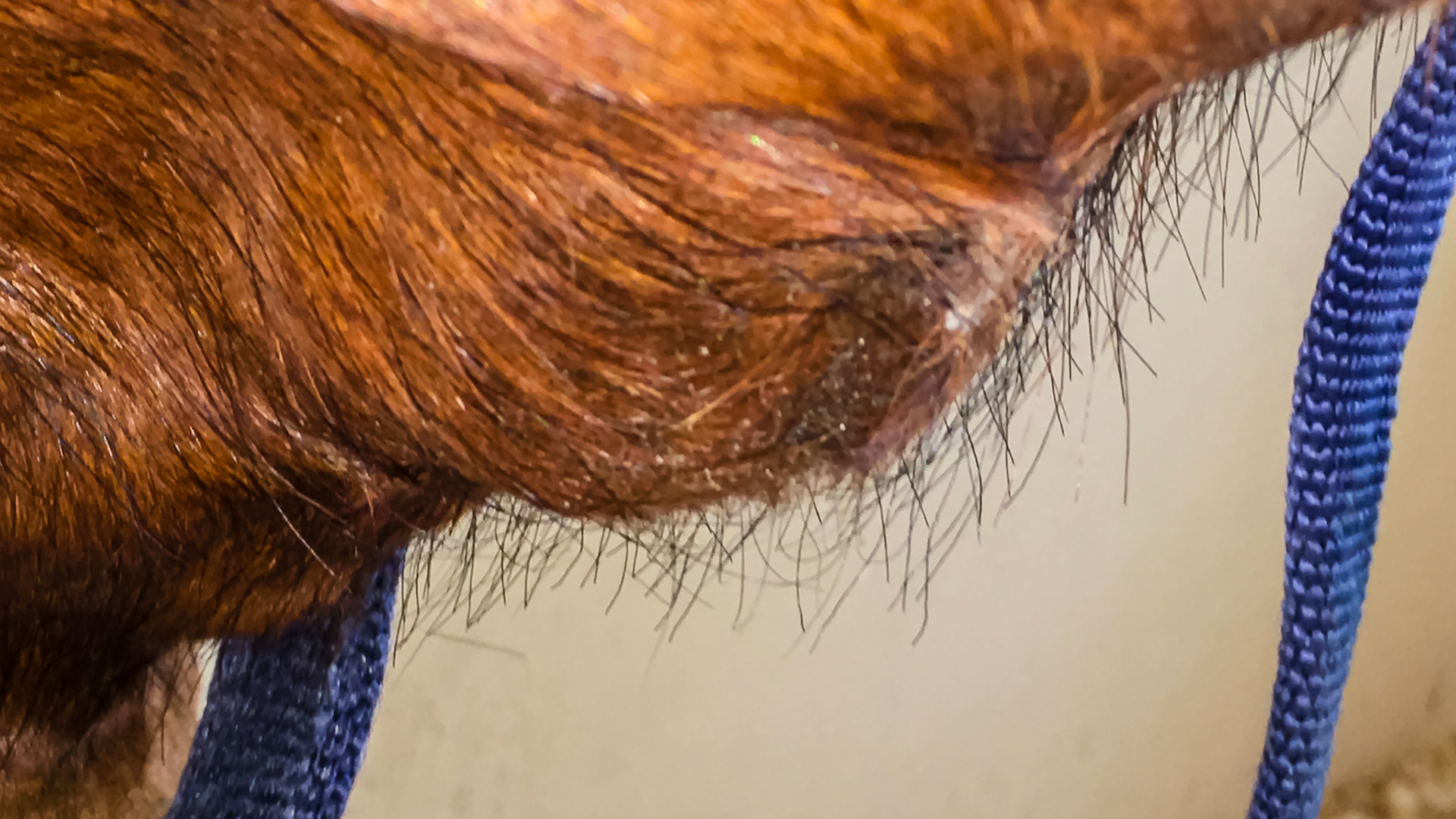
Scar from trephine extraction of molar cheek tooth #110
Teeth extracted from a 30 year old pony without drugs using cap pullers. See the movie.
Teeth extracted from a 30 year old pony without drugs using cap pullers. See the movie.
Teeth extracted from a 30 year old pony without drugs using cap pullers. See the movie.
This is cheek tooth #409 or 408 extracted from a 7 year old Hanovarian mare. There was a history of swelling followed eventually by drainage. A few antibiotics were used and finally SMZ pills were settled on. The owner tried this therapy for less than a month before she was talked into extraction. I never saw this horse. I believe that if she had waited several more months, this mare could have resolved this abscess, but owners do not want to wait or give antibiotics this long.
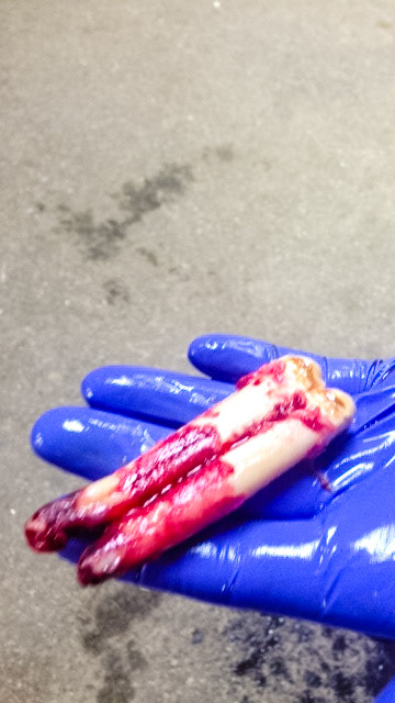
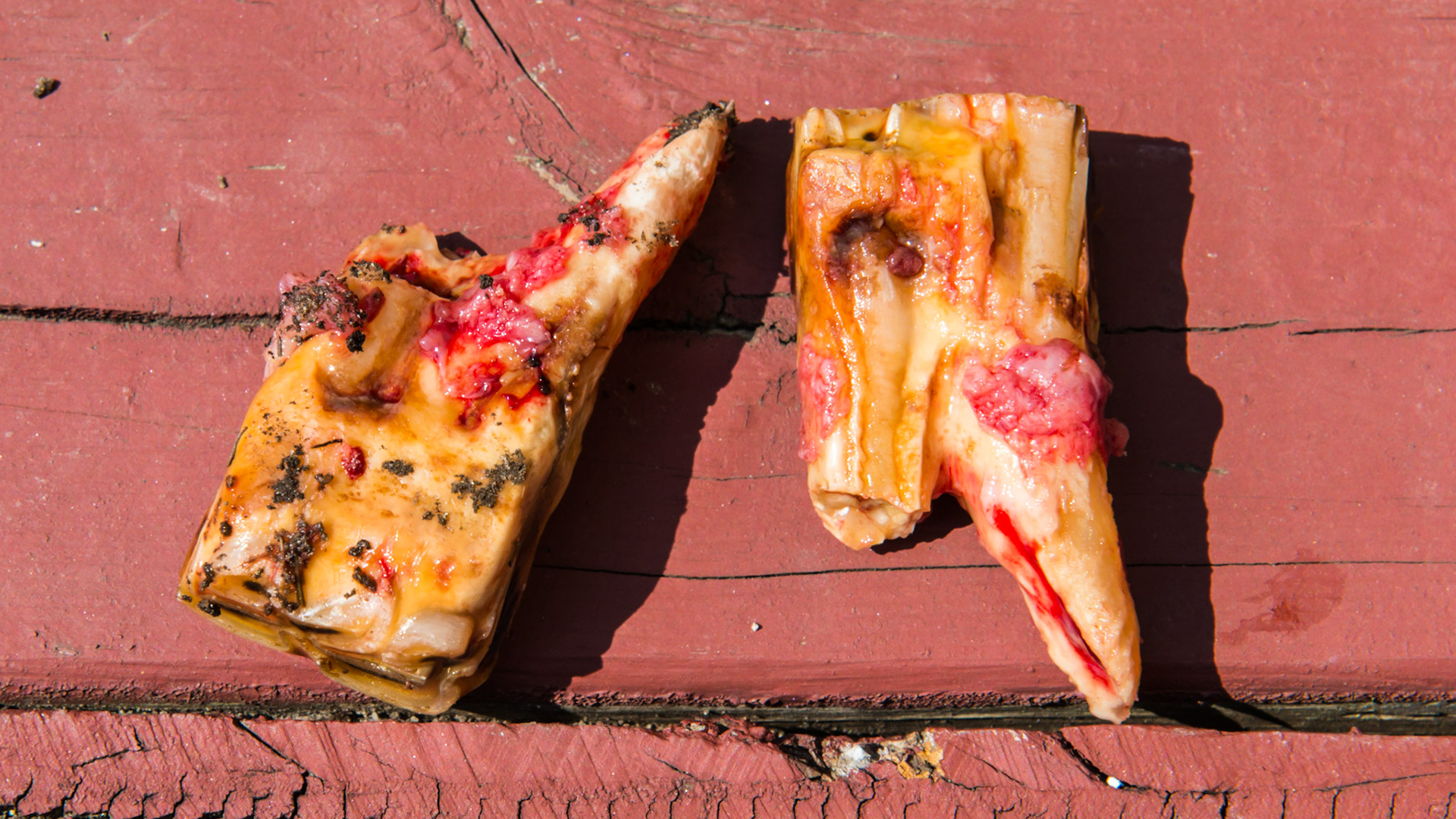
308 and 408 in a 22 year old mare. These teeth tilted laterally into the cheeks with packed hay. The tongue was unable to clean the areas. There was a malodor of decay in both teeth and both were loose and painful. Extractions were simple with IV pain medication. Both teeth had broken roots that had fractured in the past. The remaining roots were left where they were and the horse was given 3 days of Uniprim.
308 and 408 in a 22 year old mare. These teeth tilted laterally into the cheeks with packed hay. The tongue was unable to clean the areas. There was a malodor of decay in both teeth and both were loose and painful. Extractions were simple with IV pain medication. Both teeth had broken roots that had fractured in the past. The remaining roots were left where they were and the horse was given 3 days of Uniprim.
308 and 408 in a 22 year old mare. These teeth tilted laterally into the cheeks with packed hay. The tongue was unable to clean the areas. There was a malodor of decay in both teeth and both were loose and painful. Extractions were simple with IV pain medication. Both teeth had broken roots that had fractured in the past. The remaining roots were left where they were and the horse was given 3 days of Uniprim.
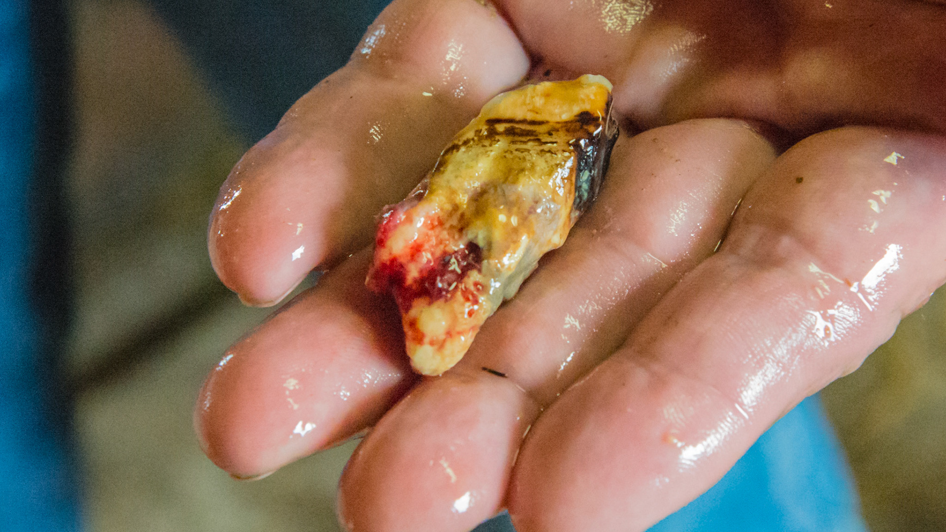
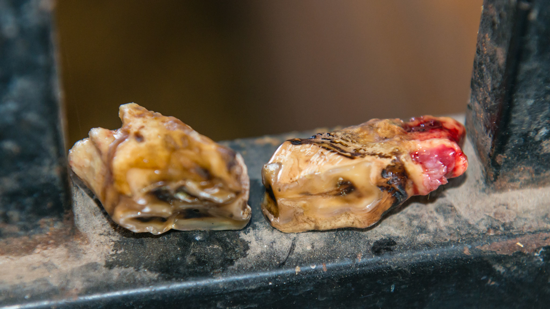
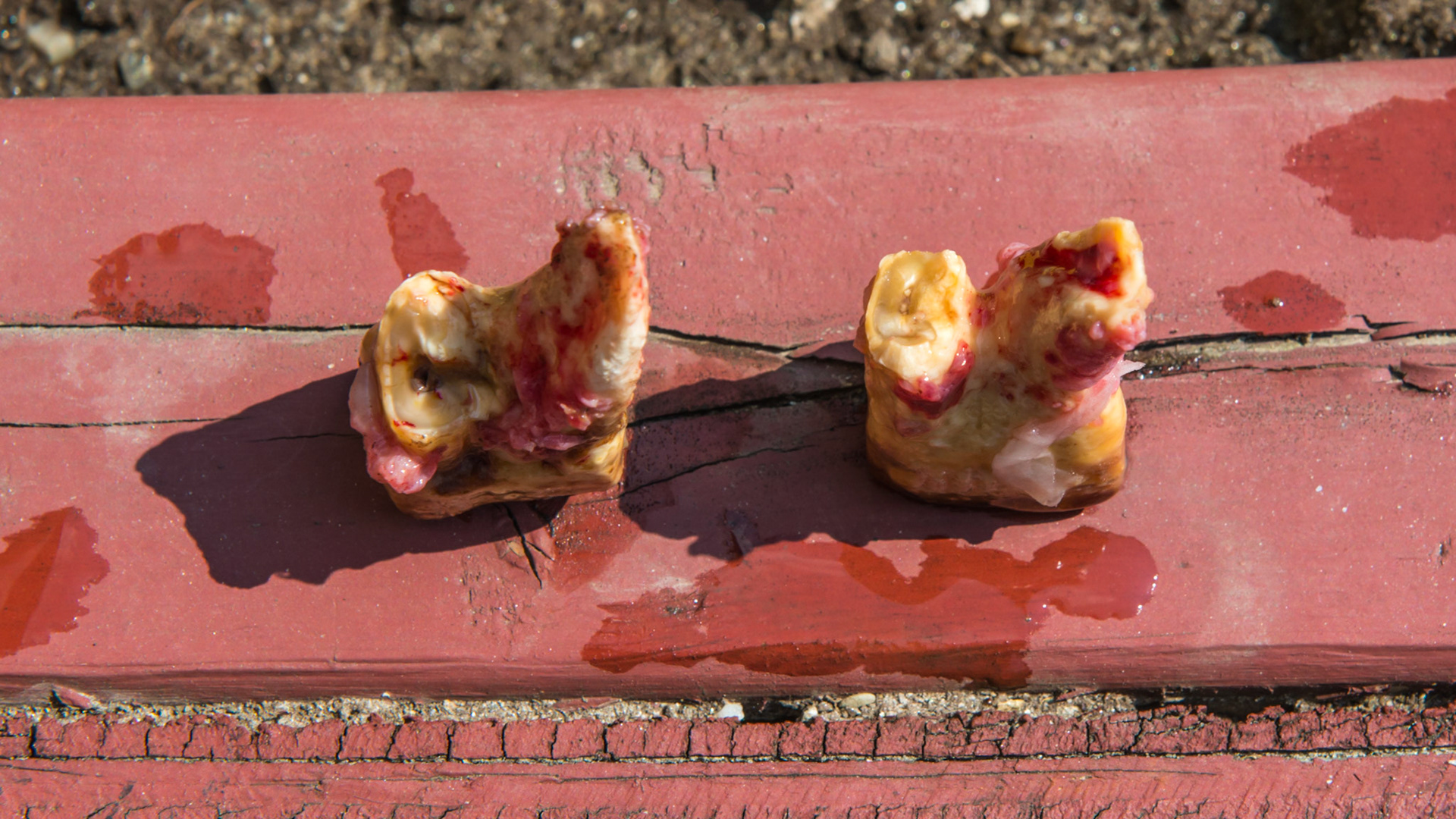
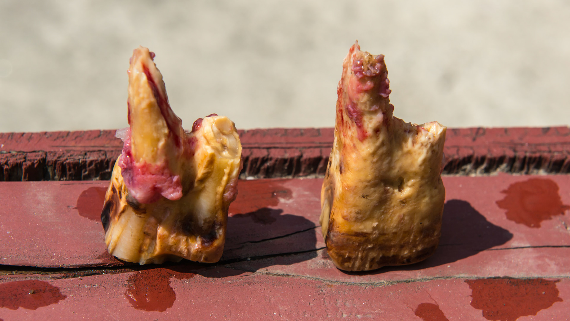
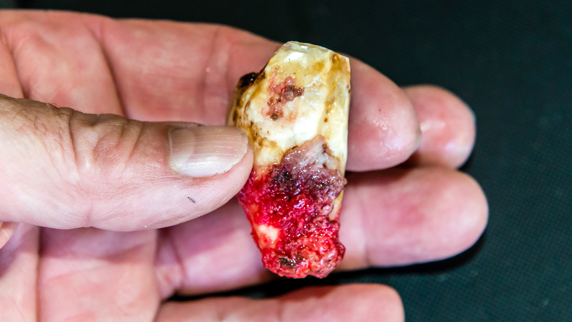
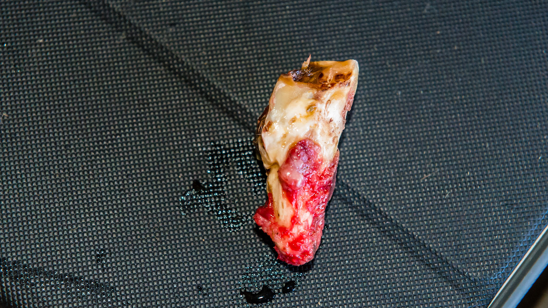
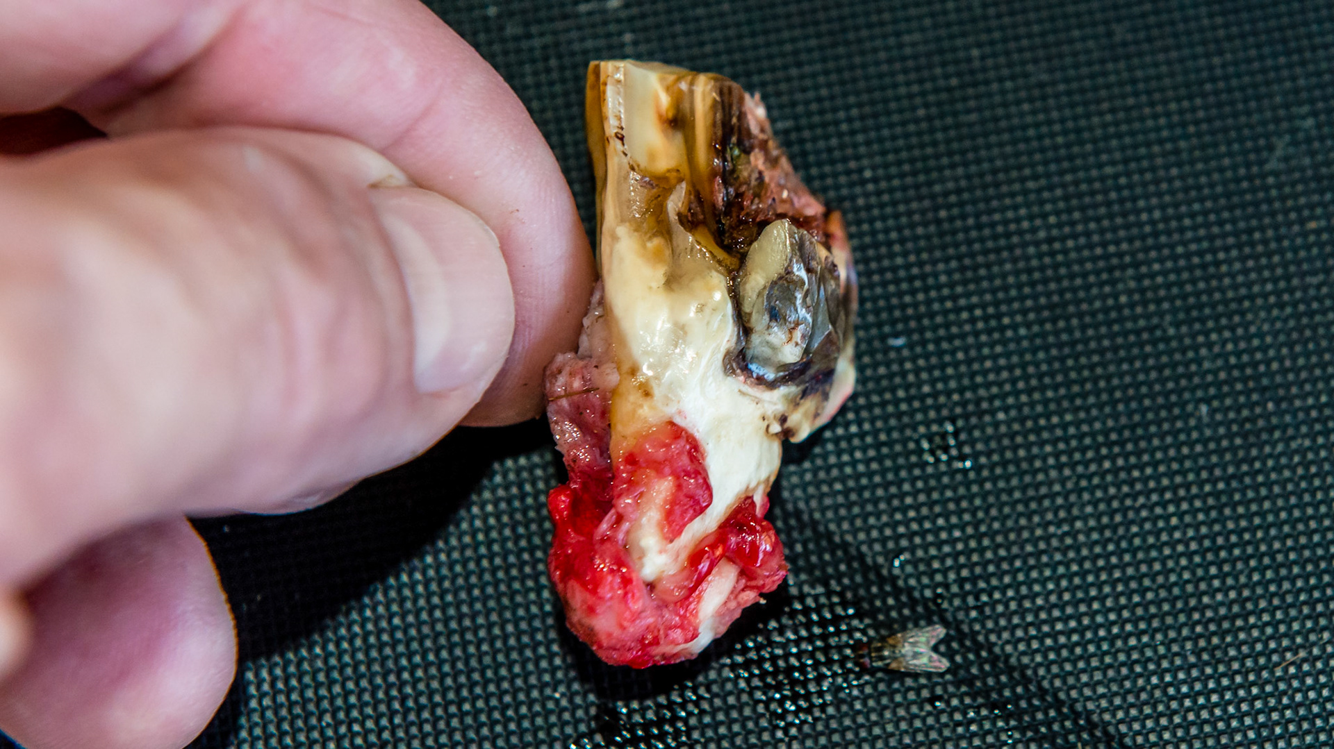
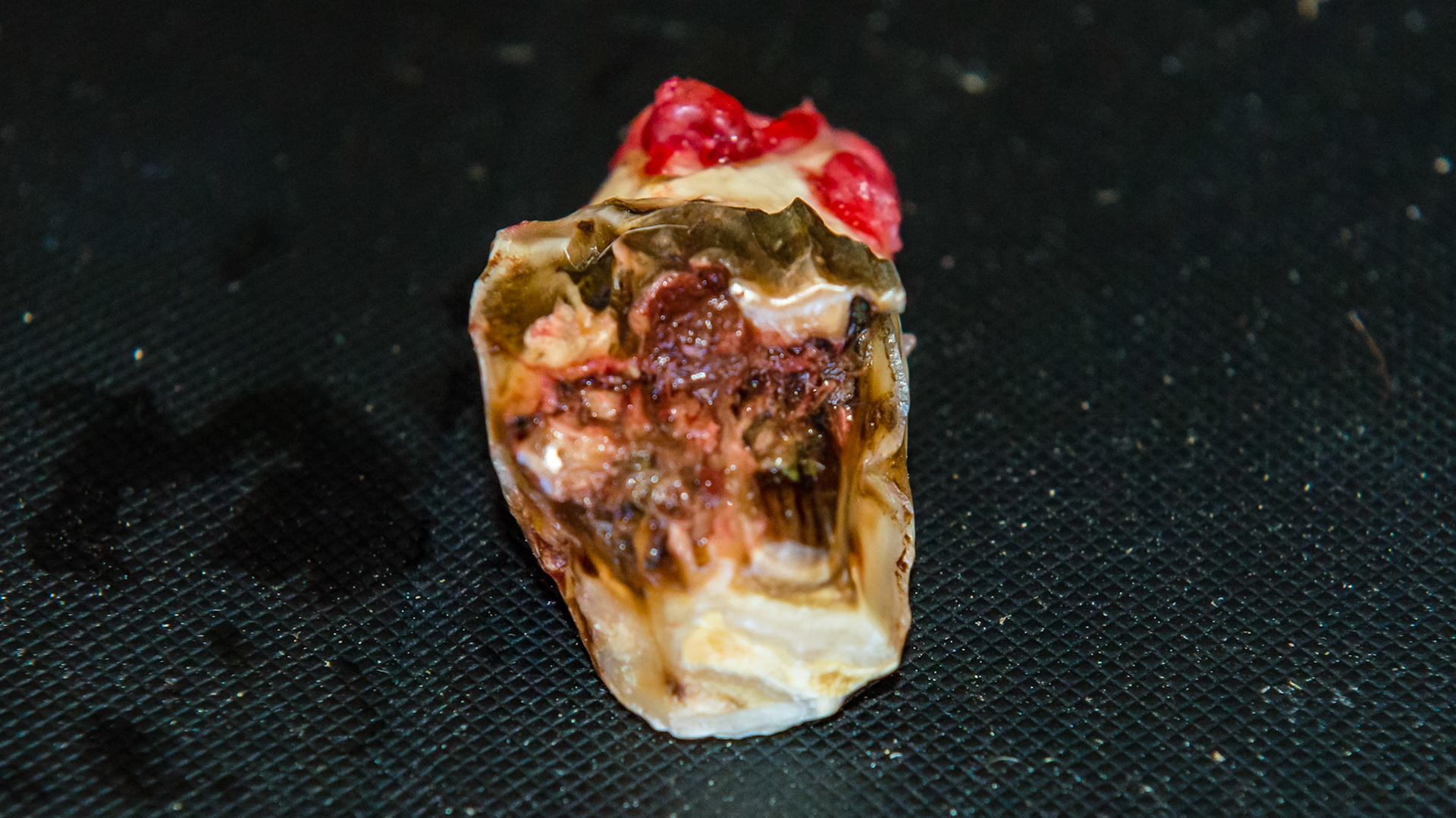
Examples of end stage teeth where the cheek tooth is held in place by only a few tufts of tissue (red). These teeth wiggle under finger pressure and are often extracted by fingers or a slight tug with forceps.
Examples of end stage teeth where the cheek tooth is held in place by only a few tufts of tissue (red). These teeth wiggle under finger pressure and are often extracted by fingers or a slight tug with forceps.
Examples of end stage teeth where the cheek tooth is held in place by only a few tufts of tissue (red). These teeth wiggle under finger pressure and are often extracted by fingers or a slight tug with forceps.
Examples of end stage teeth where the cheek tooth is held in place by only a few tufts of tissue (red). These teeth wiggle under finger pressure and are often extracted by fingers or a slight tug with forceps.
Examples of end stage teeth where the cheek tooth is held in place by only a few tufts of tissue (red). These teeth wiggle under finger pressure and are often extracted by fingers or a slight tug with forceps.
Examples of end stage teeth where the cheek tooth is held in place by only a few tufts of tissue (red). These teeth wiggle under finger pressure and are often extracted by fingers or a slight tug with forceps.
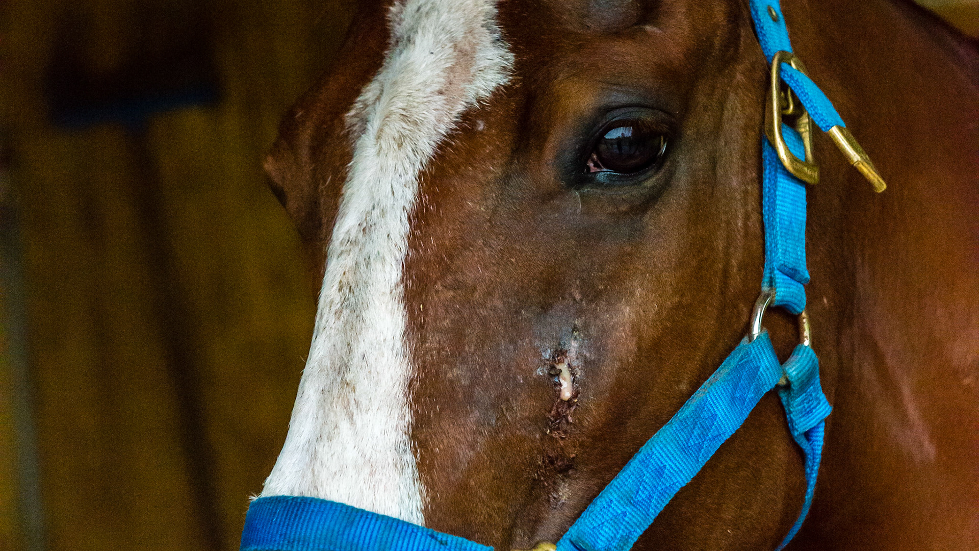
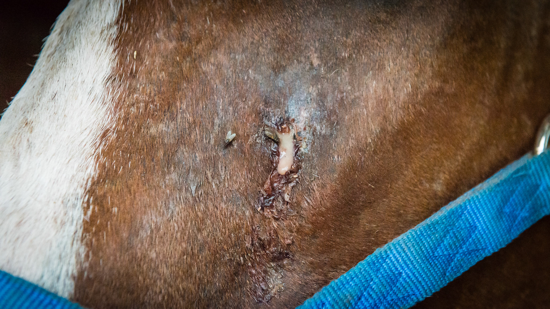
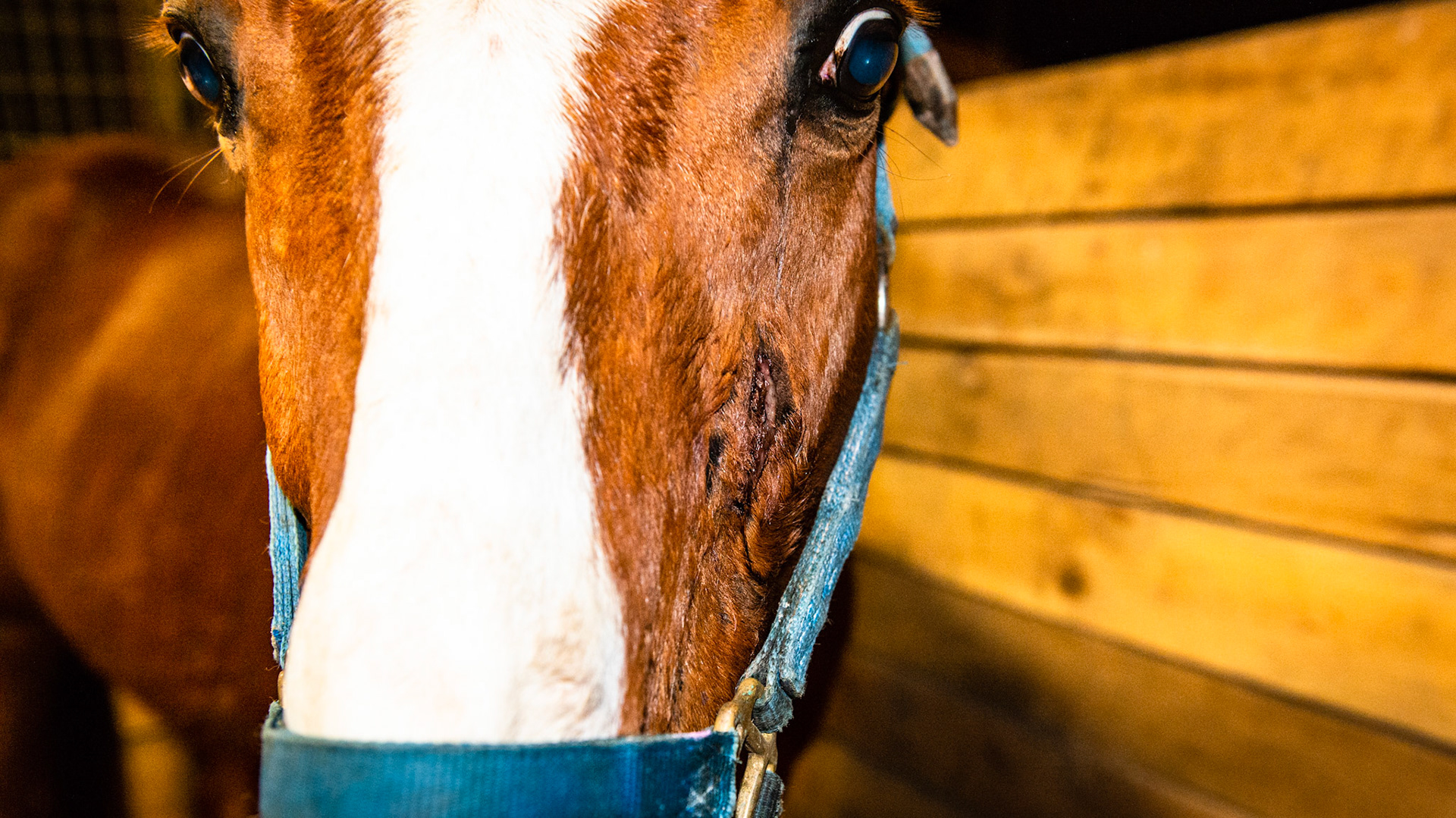
This horse has an apical tooth root abscess that has gone through the sinus and is draining through the face for almost 2 years (on and off antibiotics). The tooth was extracted but the drainage continued. About 6 to 9 months later a second adjacent tooth was extracted with no better results. The drainage continued for another year. THIS IMAGE is of the drainage before the first extraction.
This horse has an apical tooth root abscess that has gone through the sinus and is draining through the face for almost 2 years (on and off antibiotics). The tooth was extracted but the drainage continued. About 6 to 9 months later a second adjacent tooth was extracted with no better results. The drainage continued for another year. THIS IMAGE is of the drainage before the first extraction.
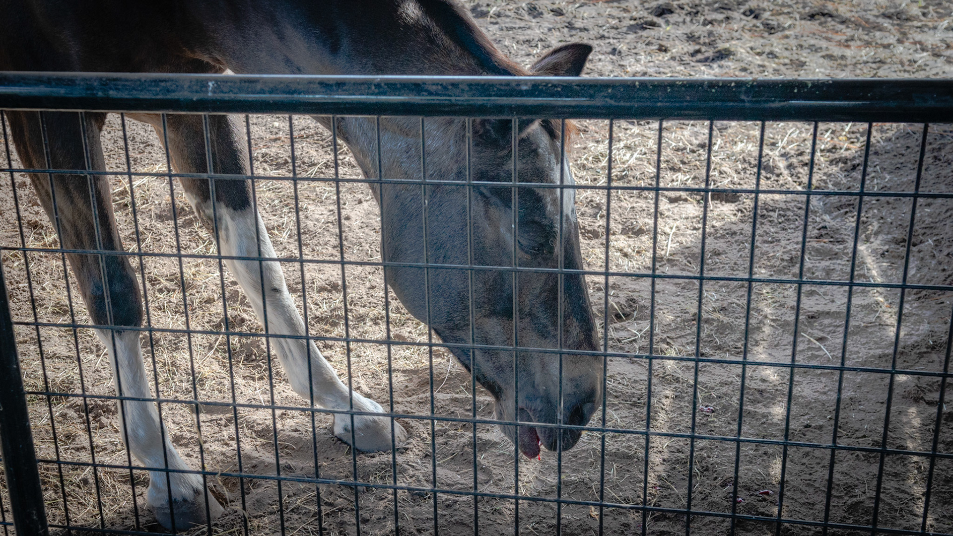
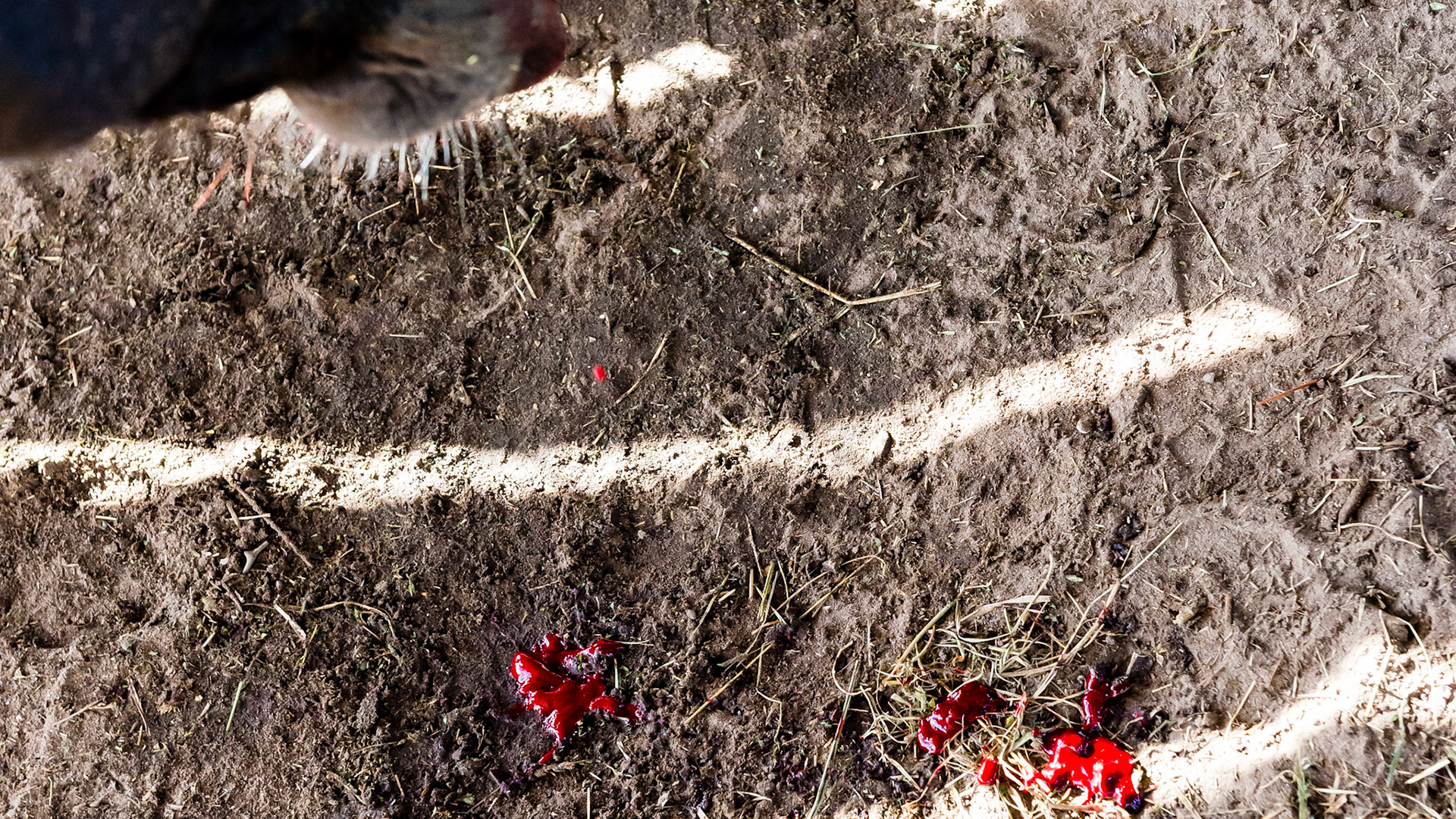
Sedated horse with a palatal sagittal fracture removal causing minor bleeding in the head down position.
Sedated horse with a palatal sagittal fracture removal causing minor bleeding in the head down position.
This lump on the ventral mandibula is hard and nonpainful. The associated tooth (308) was extracted 2 weeks earlier because of chronic draining. X-rays and a CT scan reveled infection in the roots of 308 and 310. Long term antibiotics would not resolve this. Post extraction, the drainage has stopped but the enlargement of the mandibular bone has significantly increased within the mouth while the exterior mass has organized into a taller but less round mass. Rechecking in another 4 weeks.
This lump on the ventral mandibula is hard and nonpainful. The associated tooth (308) was extracted 2 weeks earlier because of chronic draining. X-rays and a CT scan reveled infection in the roots of 308 and 310. Long term antibiotics would not resolve this. Post extraction, the drainage has stopped but the enlargement of the mandibular bone has significantly increased within the mouth while the exterior mass has organized into a taller but less round mass. Rechecking in another 4 weeks.