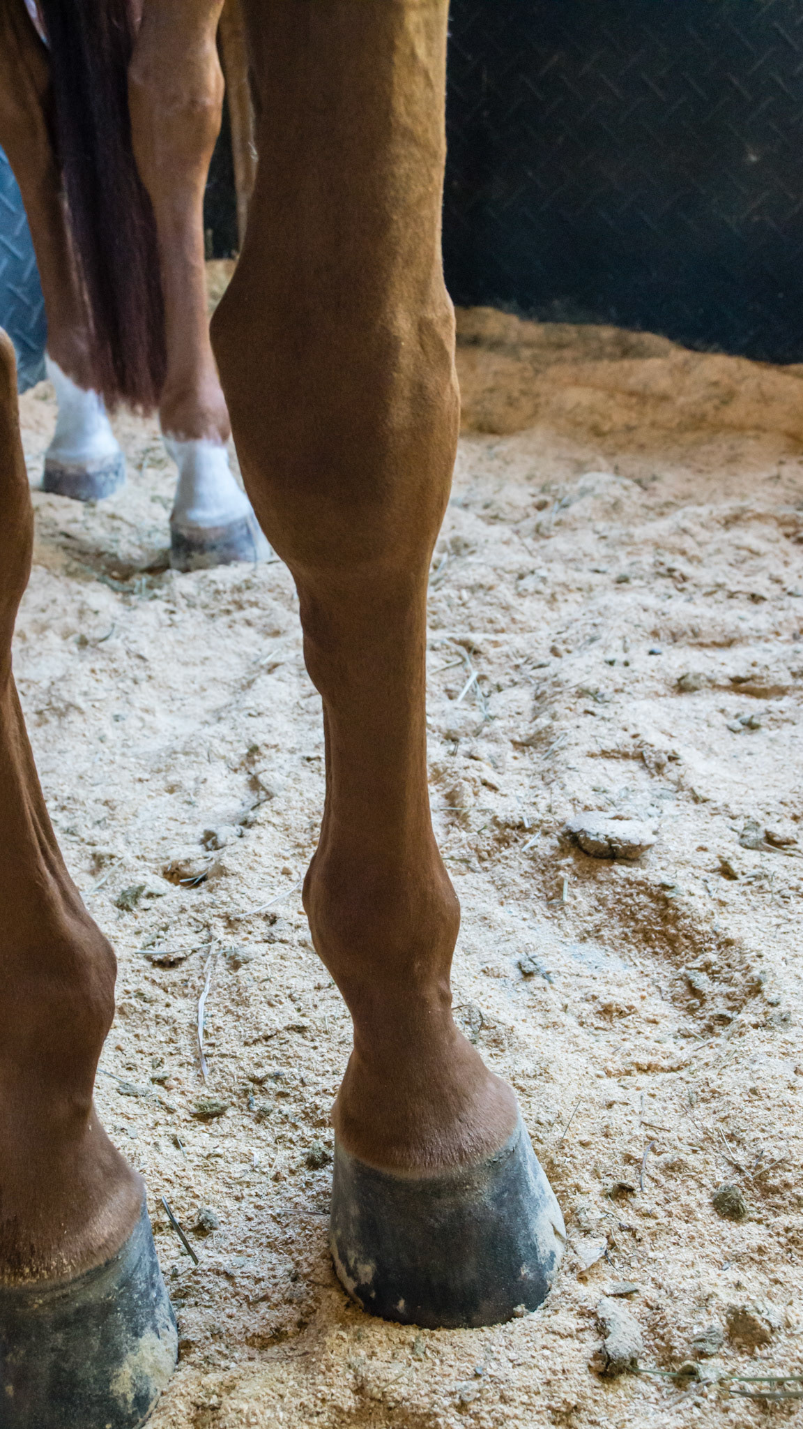
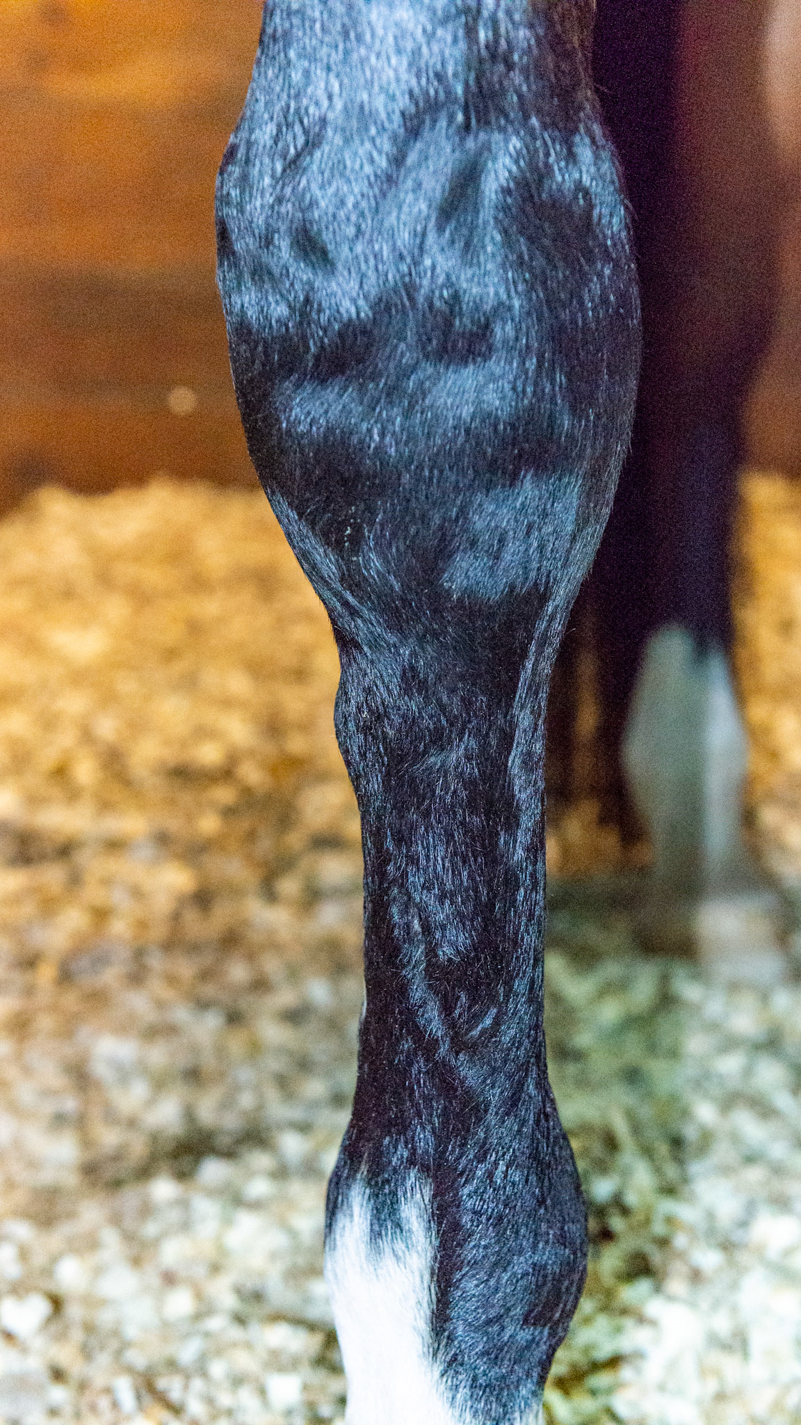
A medial splint seen on the inside of the cannon bone below the knee. This horse has bench knee conformation meaning the cannon bone is placed to the outside of the knee forming a “bench” above and to the outside of the knee forcing the horse’s weight to come down the inside of the cannon bone. This added load causes the injury. Note the more vertical inside wall of the hoof while the outside wall slightly flares out. Also note the uneven coronary band. The hoof changes are a reaction to the uneven load top the hoof caused by the bad conformation of the cannon to the forearm.
A medial splint seen on the inside of the cannon bone below the knee. This horse has bench knee conformation meaning the cannon bone is placed to the outside of the knee forming a “bench” above and to the outside of the knee forcing the horse’s weight to come down the inside of the cannon bone. This added load causes the injury. Note the more vertical inside wall of the hoof while the outside wall slightly flares out. Also note the uneven coronary band. The hoof changes are a reaction to the uneven load top the hoof caused by the bad conformation of the cannon to the forearm.
A medial splint seen on the inside of the cannon bone below the knee. This horse has bench knee conformation meaning the cannon bone is placed to the outside of the knee forming a “bench” above and to the outside of the knee forcing the horse’s weight to come down the inside of the cannon bone. This added load causes the injury. Note the more vertical inside wall of the hoof while the outside wall slightly flares out. Also note the uneven coronary band. The hoof changes are a reaction to the uneven load top the hoof caused by the bad conformation of the cannon to the forearm.
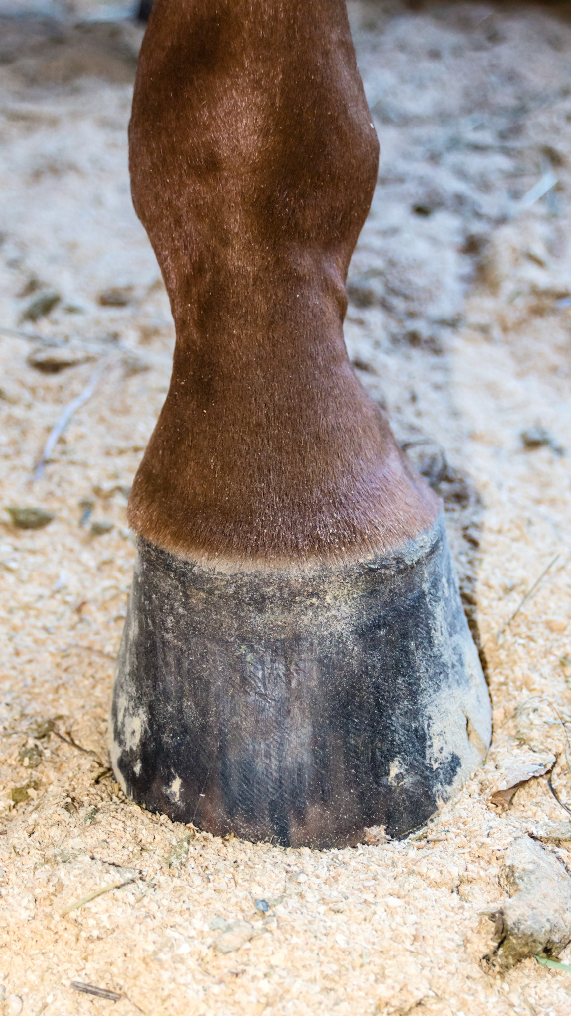
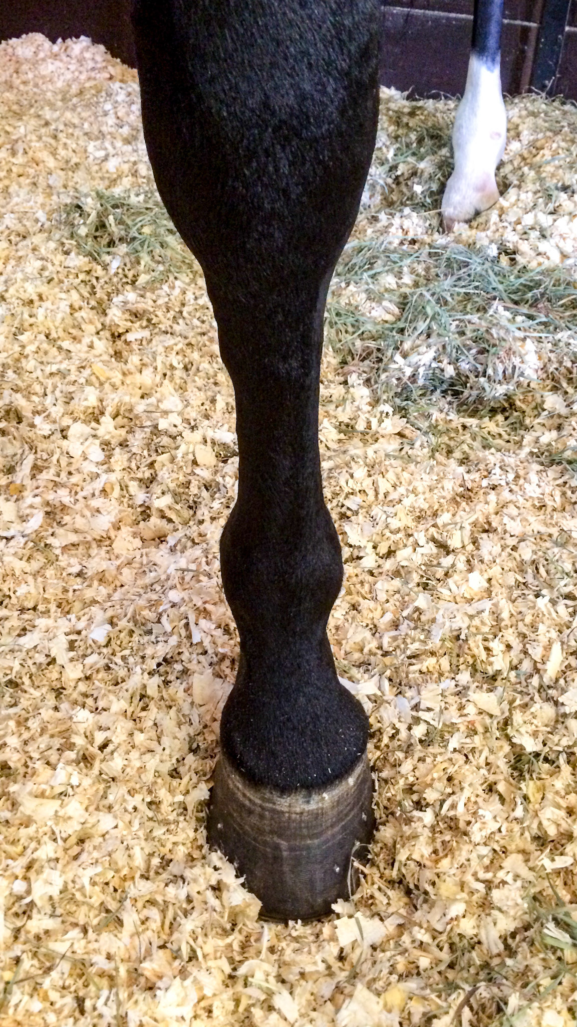
High medial splint of the LF cannon bone.
High medial splint of the LF cannon bone.
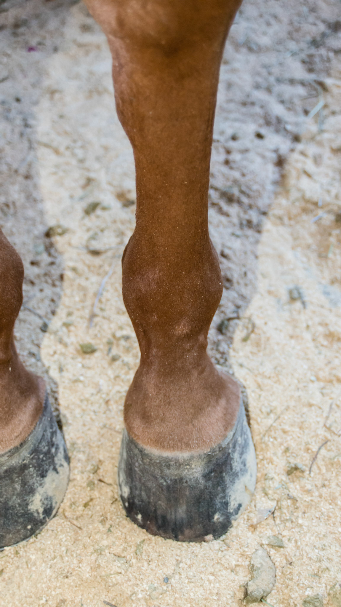
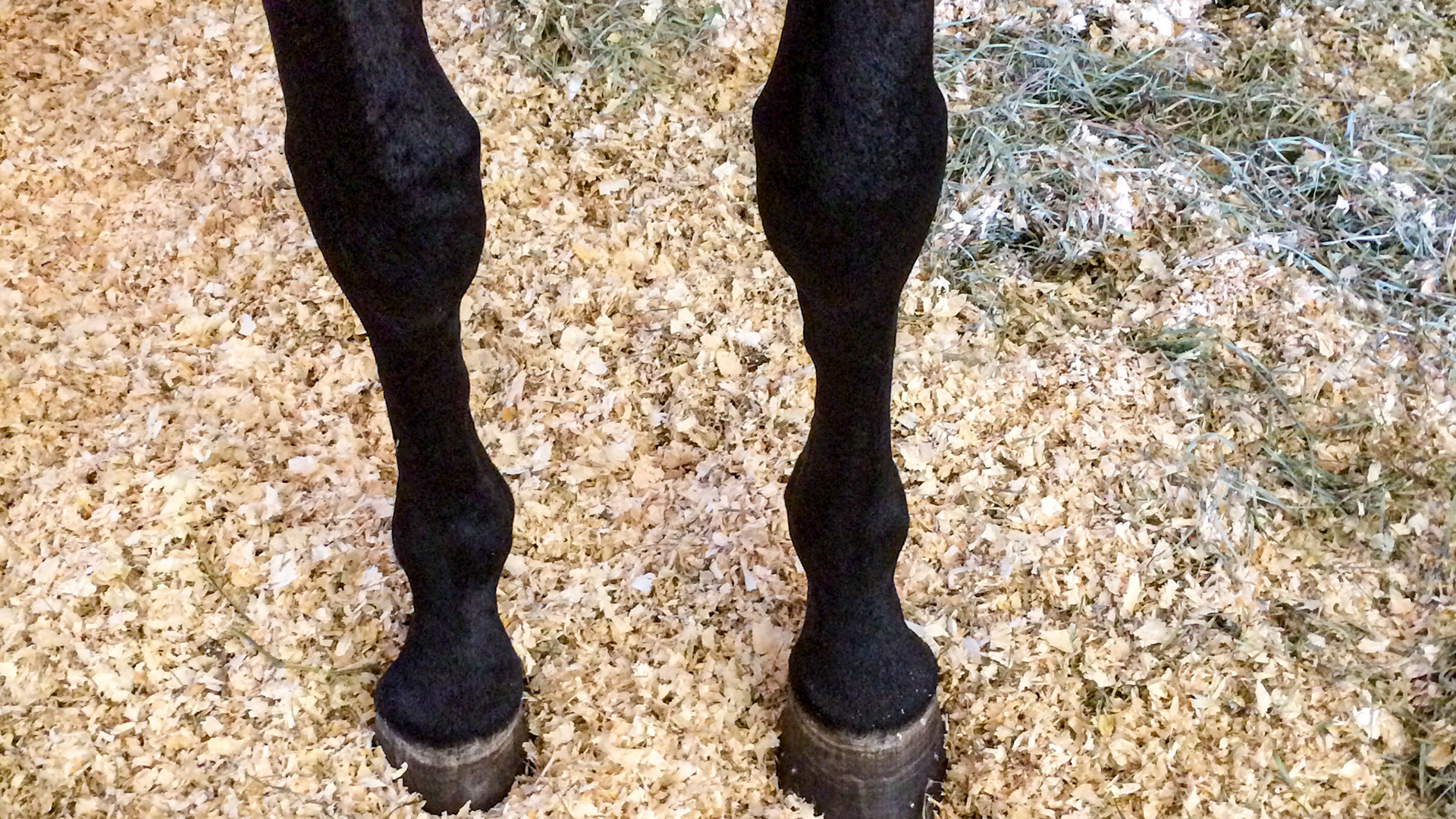
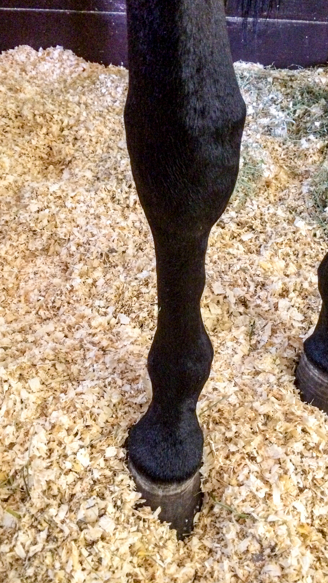
High medial splint on both cannon bones. Note the load of the horse comes down on the inside (medial side) of the cannon, fetlock, coronary band and the hoof causing the asymmetry seen in the hoof and pastern.
High medial splint on both cannon bones. Note the load of the horse comes down on the inside (medial side) of the cannon, fetlock, coronary band and the hoof causing the asymmetry seen in the hoof and pastern.
High medial splint on both cannon bones. Note the load of the horse comes down on the inside (medial side) of the cannon, fetlock, coronary band and the hoof causing the asymmetry seen in the hoof and pastern.