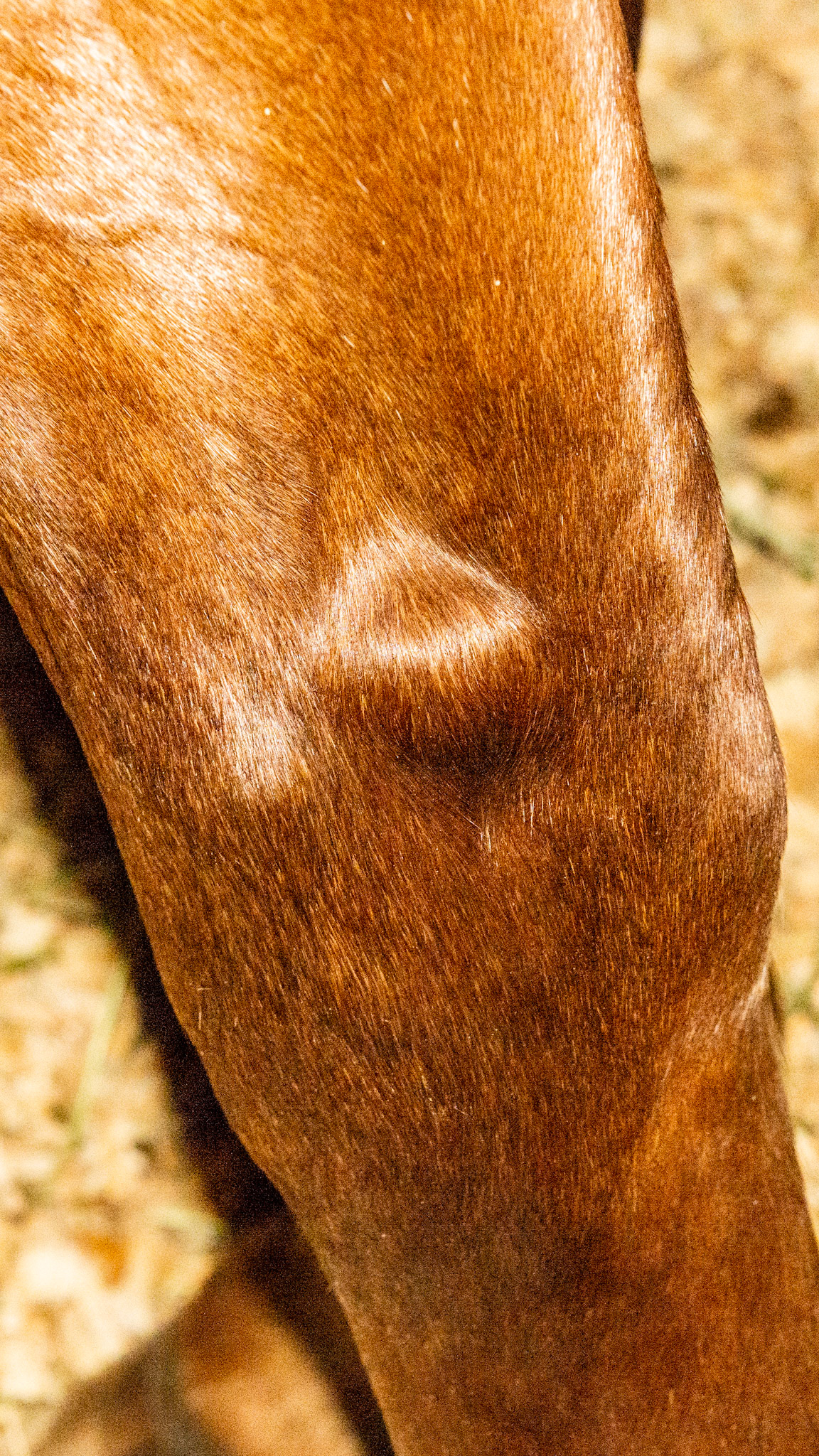
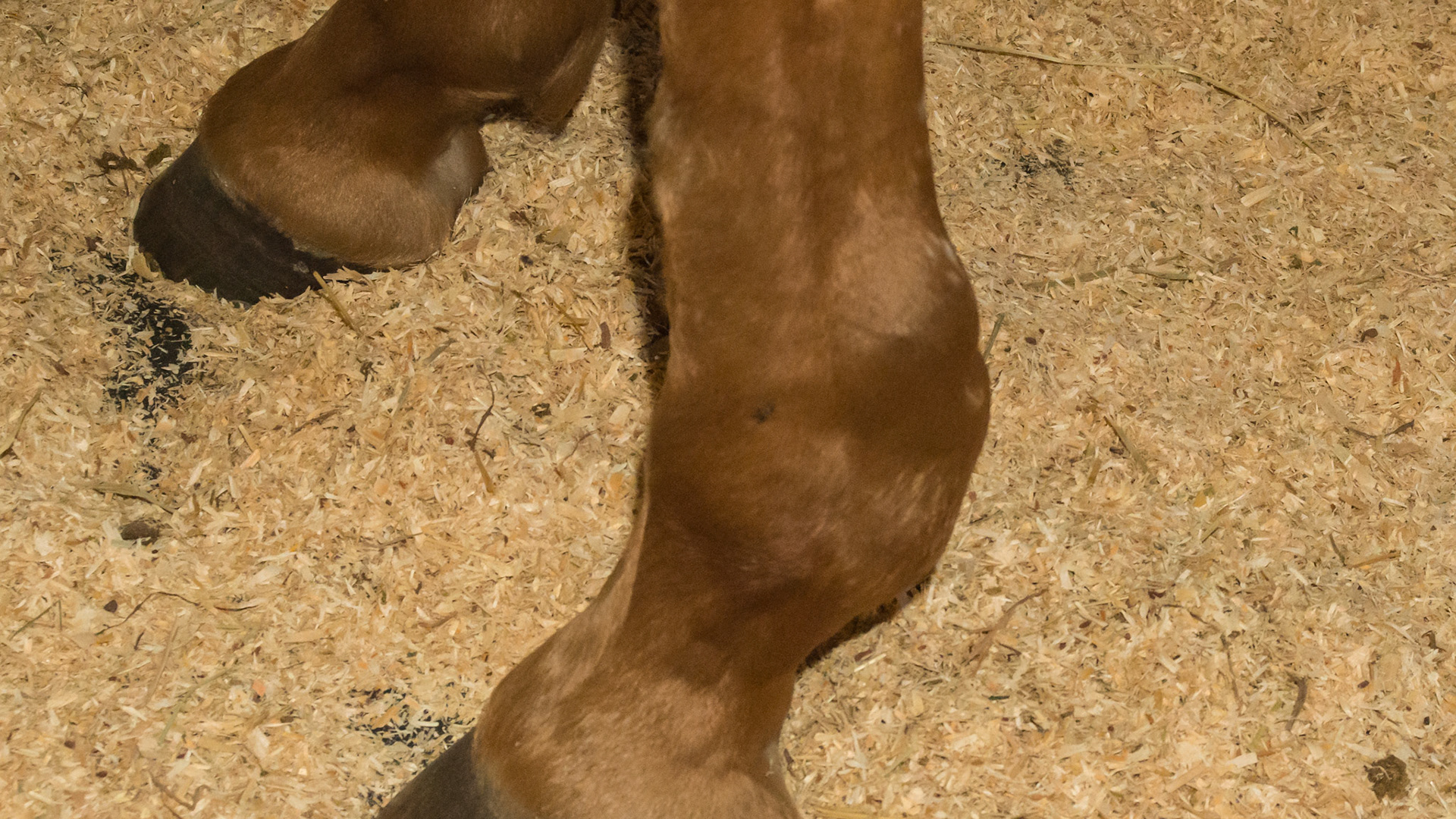
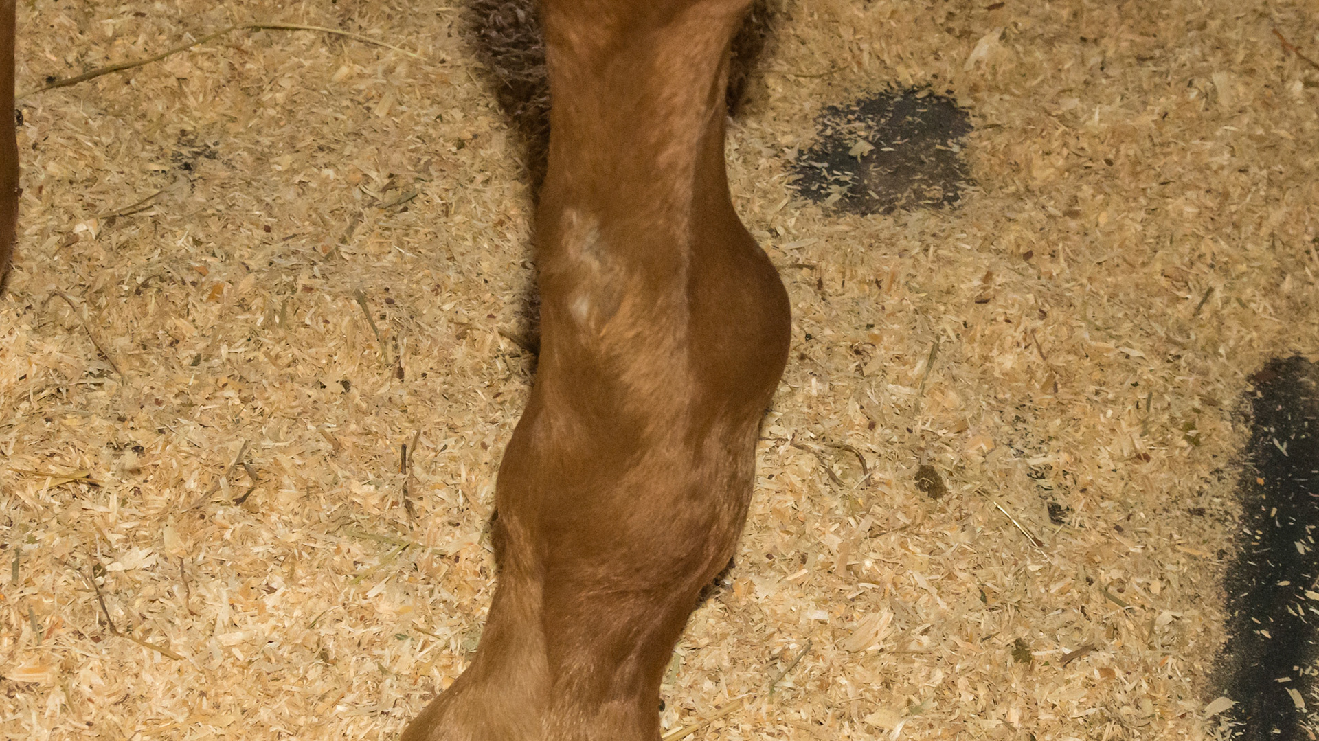
Soft fluid filled defined distention on lateral side just above the carpus (knee) of this horse.
Soft fluid filled defined distention on lateral side just above the carpus (knee) of this horse.
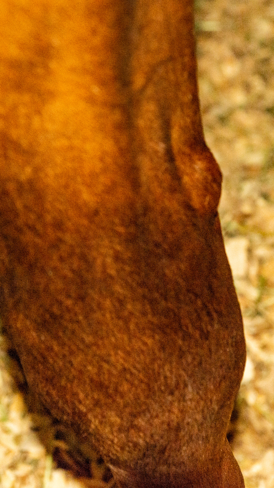
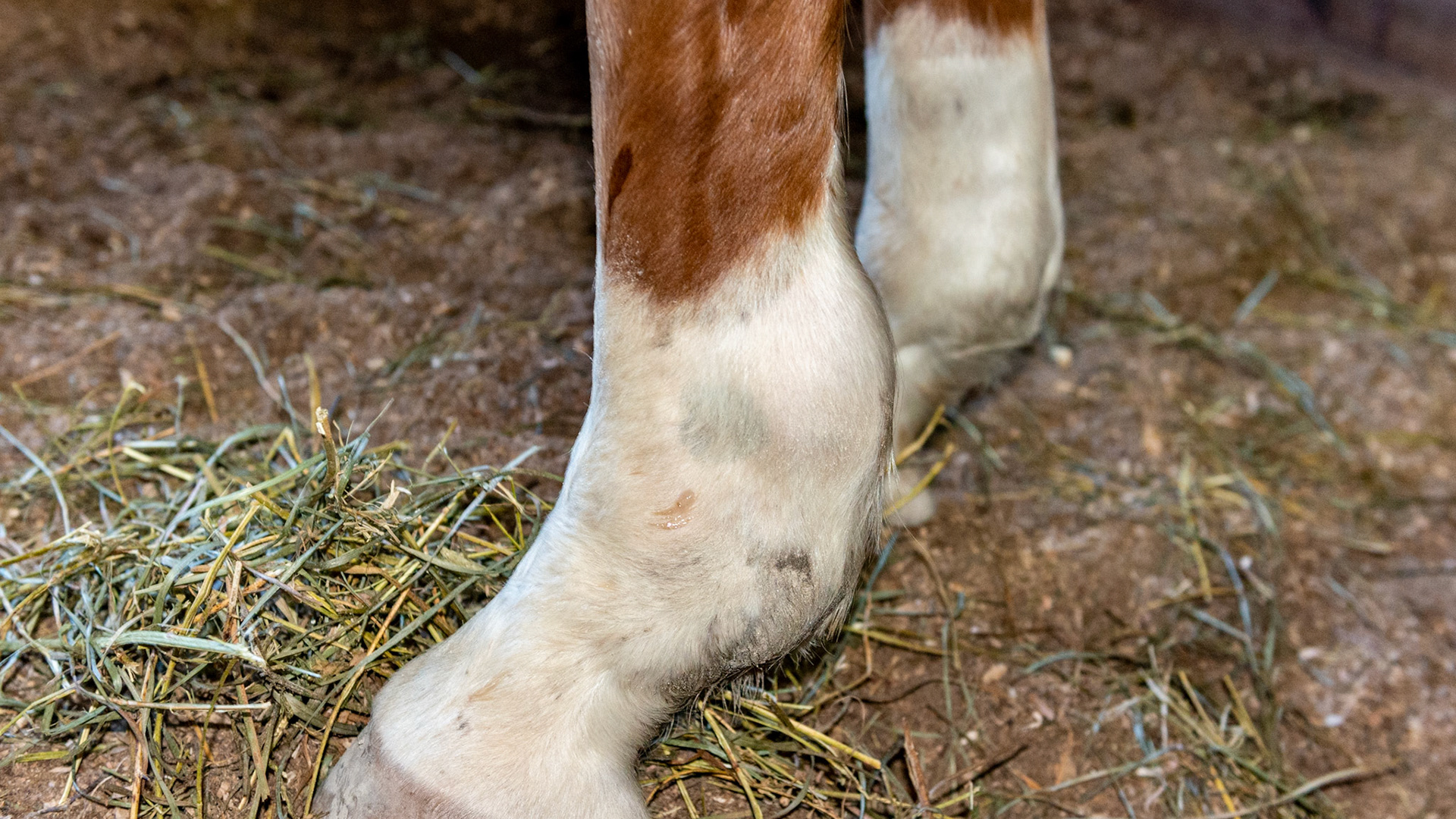
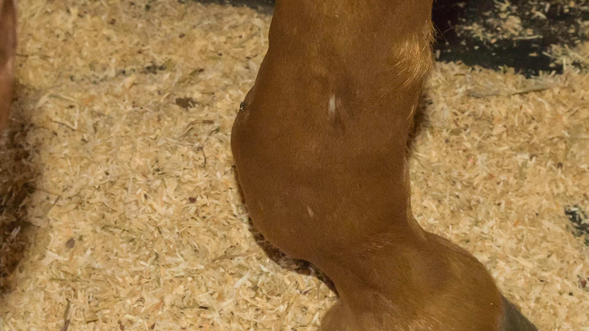
This is a severe distention of the digital tendon sheath on this forelimb. It is predominately on the outside but you can also see the flat and firm separation from the back view caused by the intact tendon dividing the sheath into medial and lateral halves. It can be differentiated from a wind puff in the side views because the bulges surround the tendon while a fetlock joint distention would be more forward of this between the cannon bone and the suspensory ligament. These are usually non-painful but are a warning that either acute or chronic trauma has occurred to the tendons at this level.
This is a severe distention of the digital tendon sheath on this forelimb. It is predominately on the outside but you can also see the flat and firm separation from the back view caused by the intact tendon dividing the sheath into medial and lateral halves. It can be differentiated from a wind puff in the side views because the bulges surround the tendon while a fetlock joint distention would be more forward of this between the cannon bone and the suspensory ligament. These are usually non-painful but are a warning that either acute or chronic trauma has occurred to the tendons at this level.
This is a severe distention of the digital tendon sheath on this forelimb. It is predominately on the outside but you can also see the flat and firm separation from the back view caused by the intact tendon dividing the sheath into medial and lateral halves. It can be differentiated from a wind puff in the side views because the bulges surround the tendon while a fetlock joint distention would be more forward of this between the cannon bone and the suspensory ligament. These are usually non-painful but are a warning that either acute or chronic trauma has occurred to the tendons at this level.
This is a severe distention of the digital tendon sheath on this forelimb. It is predominately on the outside but you can also see the flat and firm separation from the back view caused by the intact tendon dividing the sheath into medial and lateral halves. It can be differentiated from a wind puff in the side views because the bulges surround the tendon while a fetlock joint distention would be more forward of this between the cannon bone and the suspensory ligament. These are usually non-painful but are a warning that either acute or chronic trauma has occurred to the tendons at this level.
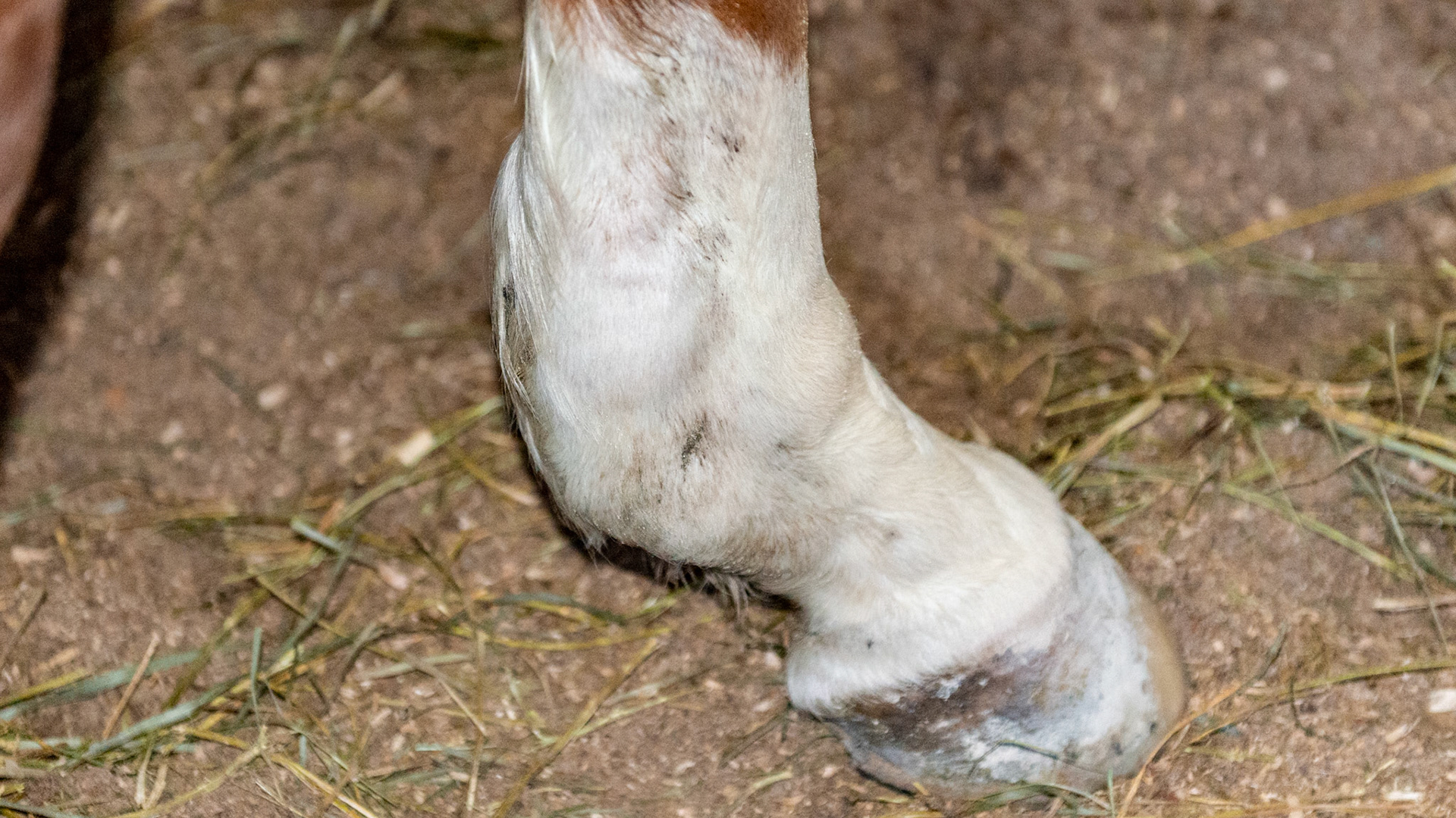
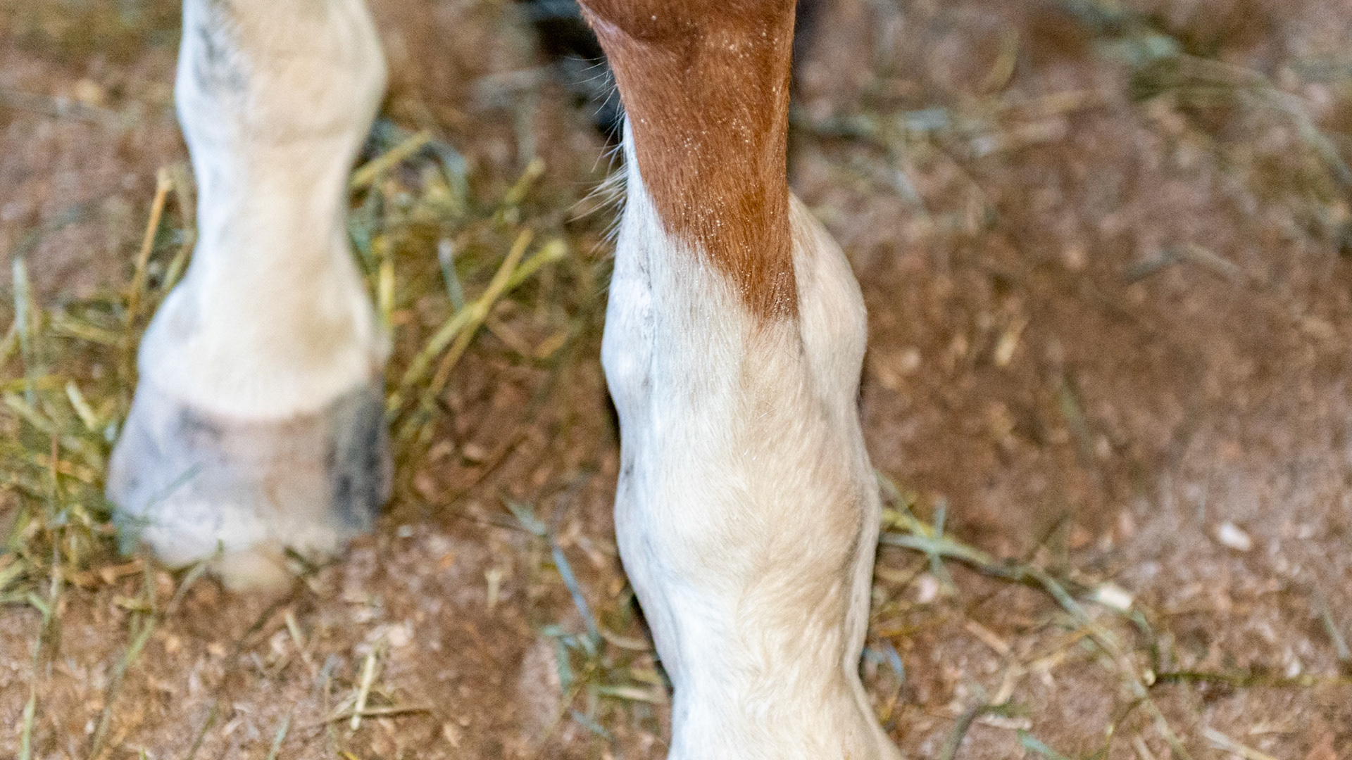
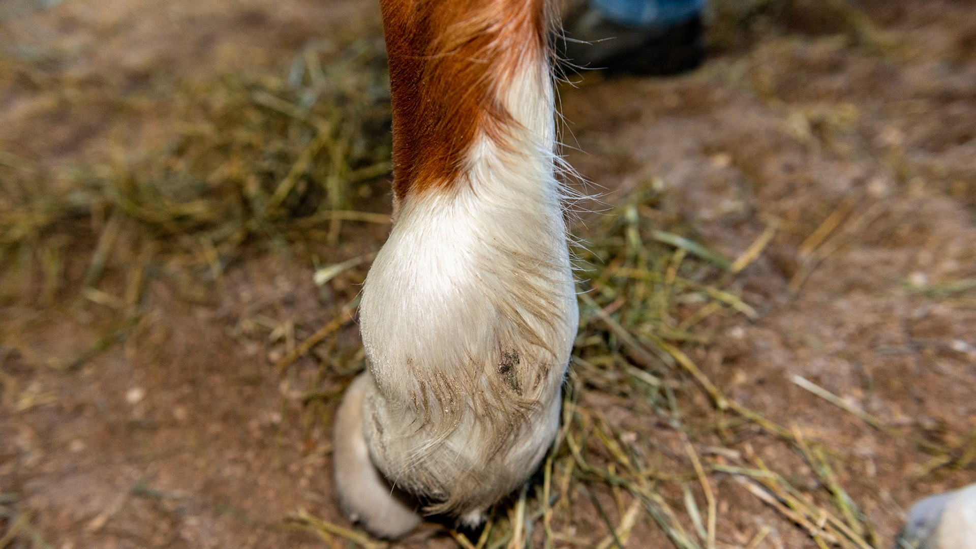
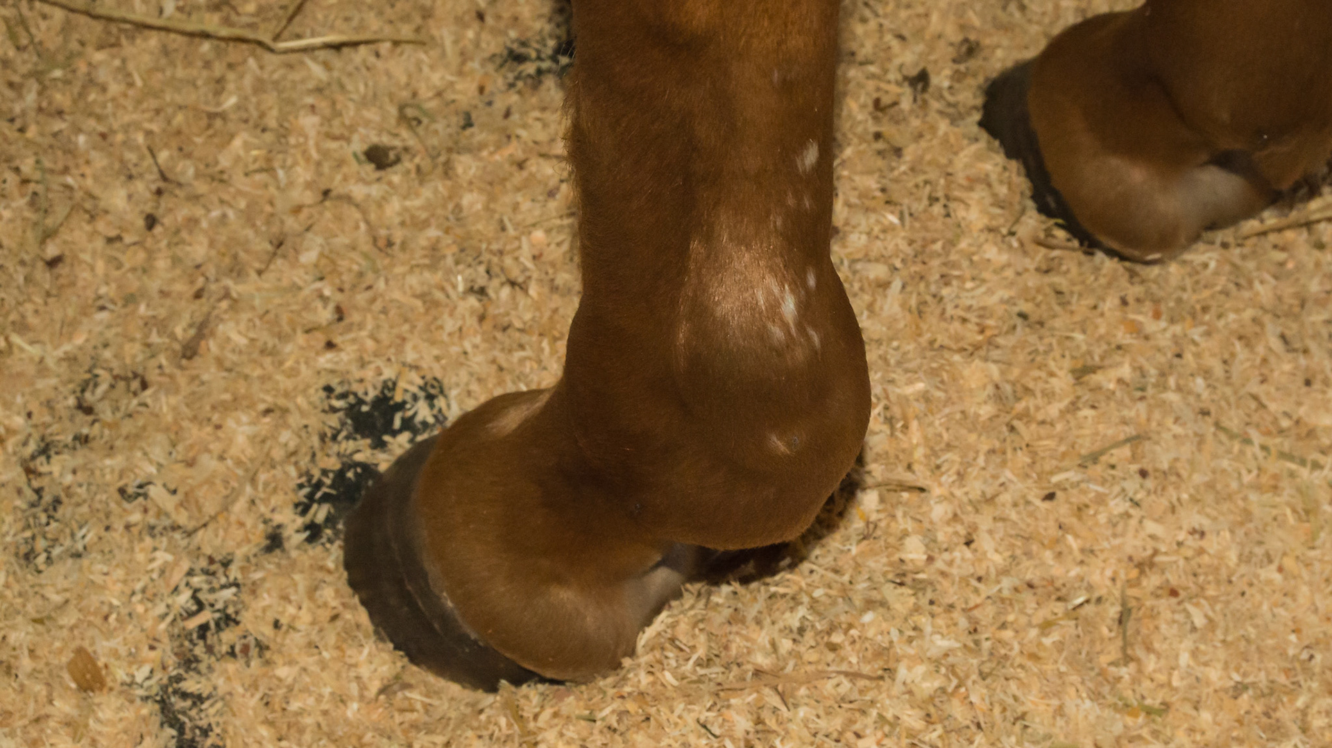
Fluid filled lateral fetlock: This is most likely a tendon sheath filling because the fluid is along the tendons, is caudal to the fetlock and is a firm fluid that can not be moved with finger pressure.
Fluid filled lateral fetlock: This is most likely a tendon sheath filling because the fluid is along the tendons, is caudal to the fetlock and is a firm fluid that can not be moved with finger pressure.
Fluid filled lateral fetlock: This is most likely a tendon sheath filling because the fluid is along the tendons, is caudal to the fetlock and is a firm fluid that can not be moved with finger pressure.
Fluid filled lateral fetlock: This is most likely a tendon sheath filling because the fluid is along the tendons, is caudal to the fetlock and is a firm fluid that can not be moved with finger pressure.